

News|Videos|March 10, 2026
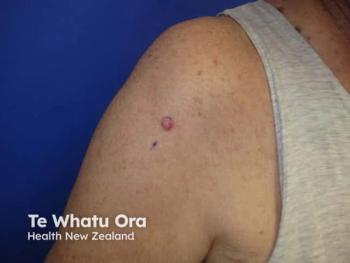
Red Light PDT Advances Field Therapy for Actinic Keratoses
Key Takeaways
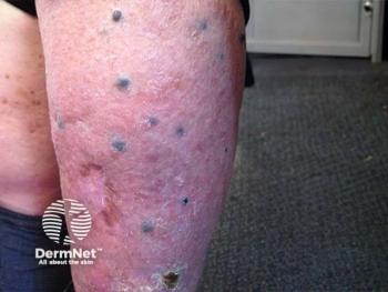
- Field-directed treatment is prioritized because a single actinic keratosis signals cumulative UV exposure and likely microscopic atypia in surrounding skin not eradicated by cryotherapy.
- Adherence limitations with 5-fluorouracil and imiquimod, driven by erythema, crusting, and erosions, make in-office PDT an attractive compliance-independent alternative.
Red light–activated photodynamic therapy offers an effective field-based option for treating both visible and subclinical actinic keratoses.
Advertisement
Episodes in this series
Photodynamic therapy (PDT) has become a cornerstone in dermatology for treating actinic keratoses (AKs), offering advantages beyond lesion-directed therapies. Anthony Rossi, MD, FAAD, FACMS, a Mohs surgeon in New York City, highlights the value of red light–activated PDT for both visible lesions and the broader areas of photodamaged skin.
Rossi emphasizes the importance of field therapy. While cryotherapy effectively removes individual AKs, it does not address the surrounding subclinical actinic damage, often referred to as field cancerization. “Even if a patient has one actinic keratosis, it’s really a marker of cumulative ultraviolet exposure,” he explained. Many additional atypical keratinocytes may exist microscopically beyond what is visible.
Topical agents such as 5-fluorouracil and imiquimod offer field-directed treatment, but adherence can be challenging due to skin reactions like erythema, crusting, or erosions. PDT, in contrast, provides a clinician-controlled alternative, reducing reliance on patient compliance.
PDT involves applying a photosensitizer, most commonly aminolevulinic acid (Ameluz; Biofrontera), which preferentially accumulates in atypical keratinocytes. Once activated by a specific wavelength of light, it induces selective cytotoxicity in dysplastic cells. Rossi often selects red light because of its deeper penetration, which is particularly useful for superficial nonmelanoma skin cancers and AKs that extend toward the basal layer of the epidermis. “Red light allows treatment of the full thickness of the epidermis, including atypical cells near the dermal-epidermal junction,” he noted.
While PDT is FDA-approved for AKs, Rossi also uses it off-label for some superficial squamous cell carcinomas and basal cell carcinomas, a practice supported by literature. Beyond cytotoxic effects, red light alone has photobiomodulation properties that can aid wound healing, reduce inflammation, and support recovery after laser procedures. Rossi also notes its use in oncology settings for inflammatory mucosal conditions.
Overall, red light–based PDT offers dermatologists a versatile approach for both visible and subclinical lesions. By combining targeted cytotoxicity with broader tissue effects, it complements surgical and topical therapies in managing AKs and chronic photodamage, reinforcing the growing emphasis on field-directed treatment strategies in clinical dermatology.
Advertisement
Related Content


Latest CME


Advertisement
Advertisement