

News|Articles|October 17, 2025
- Dermatology Times, October 2025 (Vol. 46. No. 10)
- Volume 46
- Issue 10
Bridging Personal and Clinical Experience to Better Support Patients With Breast Cancer
Author(s)Christine Ko, MD
Listen
0:00 / 0:00
Key Takeaways
- Dermatologists can be pivotal in diagnosing breast cancer, especially Paget disease of the nipple, through clinical and histopathologic findings.
- Metastatic breast cancer may present with various skin manifestations, requiring careful histopathologic evaluation for accurate diagnosis.
Explore essential insights on breast cancer diagnosis and dermatology, highlighting key practices for early detection and patient care.
Advertisement
As a medical student, there was a period of time when I had a minor form of nosophobia,1 preoccupied with the belief that I had X or Y diagnosis as I learned about each one. As for many medical students, this preoccupation with my own symptoms and health status (or lack thereof) subsided as diseases and illness scripts became more familiar. I have been blessed with good overall health for most of my life, with nary a broken bone or surgery other than a cesarean section for the birth of my first child. The potential for personal illness receded in my mind to become not quite an impossibility but something that I was perhaps immune to, as if somehow diagnosing patients with various ailments granted immunity via desensitization, akin to allergy shots.
So, it was with a certain amount of wonder that I felt a roundish lump slightly below my right armpit area several days after my birthday in 2022. It felt like an epidermoid cyst. Although it was new, it almost seemed superficial enough that perhaps I could even cut it out myself—mere “skin surgery.” For about a week, I intermittently mulled over the next step until I impulsively called my primary care physician, who told me to inform my obstetrician-gynecologist (ob-gyn). Not having seen anyone regularly in ob-gyn, I was scheduled with someone unfamiliar to me, a nurse practitioner who brusquely suggested that the lump was unlikely to be anything serious. She commented that I was too young to have breast cancer, which made no sense to me medically. Ultimately, though, she somewhat reluctantly ordered a mammogram, giving me the number to make an appointment.
Although the mammogram was not definitive, a biopsy was recommended and scheduled for a week later. I was called with an initial pathology result indicating ductal carcinoma of the breast, about 4 hours after the biopsy had been done—a very quick turnaround time. In light of my experience, and in honor of Breast Cancer Awareness Month, here are a few general practice points for all dermatologists, as well as a few points to ponder.
General practice points for all dermatologists
Paget Disease of the Nipple
Clinical and histopathologic findings
Dermatologists may be the first to diagnose a primary breast carcinoma, particularly for women who present with a nipple lesion. Common presentations include unilateral nipple erythema, erosion, weeping/crusting, or even bleeding.2 Histopathologic findings of Paget disease of the nipple include atypical cells with prominent cytoplasm, singly and in clusters that may appear to surround ductal lumina, scattered within the epidermis. These cells are generally cytokeratin 7 (CK7) positive.2 Pagetoid squamous cell carcinoma in situ of the nipple can have similar atypical Pagetoid cells, but immunohistochemistry can be helpful in distinguishing these 2 entities.3 Typically, Paget disease of the nipple is positive for CK7 and CAM 5.2, whereas Bowen disease of the nipple is p63 positive.3
Metastatic Breast Cancer
Clinical findings
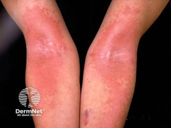
Metastatic breast carcinoma may present as erythematous papules (Figures 1 and 2) or patch(es)/plaque(s) on the breast or trunk (that may mimic erysipelas, termed inflammatory carcinoma or carcinoma erysipeloides), alopecia of the scalp, carcinoma en cuirasse (ie, indurated skin, with prominent follicular openings), or telangiectasias.4 The surface of lesions may be crusted or eroded.
Histopathologic findings
There are various histopathologic patterns of metastatic breast cancer.4 Atypical cells may present as single units between collagen bundles (Figure 3), a pattern more typical of metastatic lobular carcinoma of the breast. Or, especially for metastatic ductal carcinoma of the breast, atypical cells may cluster in epithelial islands, cords, or sheets with ductal/glandular differentiation (Figure 4). Carcinoma cells can be solely present within vascular spaces, and such involvement within vessels may only be identified with multiple tissue sections. For metastatic mucinous carcinoma of the breast, epithelial islands float in pools of mucin.
Typically, most breast carcinomas are positive with CK7 and negative with CK20. Metastatic adenocarcinomas of the breast may occasionally mimic a benign adnexal tumor, with the latter generally being diffusely positive with CK5/6 as well as p40 or p63.5 For treatment purposes, labeling for HER2, estrogen receptor, and progesterone receptor is generally performed.
Tips for Dermatologists on Sampling
For Paget disease of the nipple and metastatic breast carcinoma, a punch or wedge biopsy is often preferred over a shave biopsy, as a shave biopsy may be inadequate to assess for any underlying carcinoma. In greater than 90% of cases of Paget disease of the nipple, there is associated in situ or invasive ductal carcinoma.6
A Few Things to Ponder
21st Century Cures Act vs Physician-Supplied Diagnosis: How Quickly Does a Patient Want to Know Their Diagnosis?
As I shared in my personal story, I received my diagnosis via a phone call from the radiologist who did the ultrasound-guided biopsy, 4 hours after the procedure. I would say that is fast, and I believe it was amazingly courteous and kind of the physician to call me right away. That being said, it is not always possible for a physician to call a patient immediately, right when results are posted in an electronic medical record. And in such a case where there may be a time delay, my personal preference would be to know right away.
Even though the radiologist called me after only 4 hours had passed since my procedure, she had told me that she would call me as soon as possible and that biopsy results might be ready within 2 hours. After 2 hours, I was anxiously checking my electronic record for results, refreshing the screen every 10 minutes or so. When the radiologist called me, the diagnosis was not yet posted in my chart, as it was only preliminary, and stains had not yet been performed on the tissue sample.
Despite my personal preference regarding my breast cancer diagnosis, I have learned through seeing patients that not every patient has the same opinion as I do. Some only want to hear results via a physician. Some are like me and want to know the results right away. When I do a biopsy, I have come to realize that I need to ask patients: Are you OK with seeing the result before I am able to call you? Then we can talk about it and decide on a plan.
Uninterpretable Reports, Even as a Physician
Despite my subspecialty being related to pathology, my final pathology report was not easily interpretable to me. A friend of mine connected me to a well-known breast pathologist, who kindly spent 40 minutes on the phone with me, explaining all the nuances of the report, such as the Breast Imaging-Reporting and Data System and the prognostic significance of perineural invasion, as well as grading. I am grateful to that pathologist. Every patient does not have a personal connection to a pathologist who can explain all the words in a complex report, and time, care, and patience during scheduled visits are invaluable to patients.
Caring Physicians Are Needed Now More Than Ever
Overall, I am inexpressibly grateful to the physicians who comprise the team that took care of me, the nurse practitioner, the radiologist, the medical, surgical, and radiation oncologists. I feel very lucky to have had physicians who were and are willing to spend time with me, listen to my concerns, and give me answers to various questions. One in 7 women will get breast cancer, and I hope this article might help these patients and their physicians in some way.
Christine Ko, MD, is a professor of dermatology and pathology at Yale School of Medicine in New Haven, Connecticut.
References
- Hunter RC, Lohrenz JG, Schwartzman AE. Nosophobia and hypochondriasis in medical students. J Nerv Ment Dis. 1964;139:147-152. doi:10.1097/00005053-196408000-00008
- Alkul M, Lin CP, Truitt J, Tarbox MB. A tale of three common nipple diseases. Proc (Bayl Univ Med Cent). 2022;35(3):354-356. doi:10/1080/08998280.2022.2027195
- Wieland R, Adhikari P, North J. The utility of p63, CK7, and CA5.2 staining in differentiating pagetoid intraepidermal carcinomas. J Cutan Pathol. 2023;50(12):1110-1115. doi:10.1111/cup.14446
- Hussein MR. Skin metastasis: a pathologist’s perspective. J Cutan Pathol. 2010;37(9):e1-e20. doi:10.1111/j.1600-0560.2009.01469.x
- Qureshi HS, Ormsby AH, Lee MW, Zarbo RJ, Ma CK. The diagnostic utility of p63, CK5/6, CK 7, and CK 20 in distinguishing primary cutaneous adnexal neoplasms from metastatic carcinomas. J Cutan Pathol. 2004;31(2):145-152. doi:10.1111/j.0303-6987.2004.00147.x
- Jang N, Kang S, Bae YK. Mammary Paget’s disease without underlying malignancy of the breast. Yeungnam Univ J Med. 2018;35(1):99-103. doi:10.12701/yujm.2018.35.1.99›
Articles in this issue
9 months ago
Cosmetic Trends of Dermatologic Interest in 202510 months ago
Identifying Breast Issues Beyond the SkinAdvertisement
Related Content
Advertisement


Latest CME


Advertisement
Advertisement
Trending on Dermatology Times
1
Tapinarof Simplifies Long-Term Atopic Dermatitis Treatment Regimens
2
Sanofi Discontinues Development of OX40L Inhibitor Amlitelimab in Atopic Dermatitis
3
The Aesthetic Edge: July 2026
4
SPD 2026: Phase 3 Success for Lebrikizumab in Younger Pediatric AD Patients
5