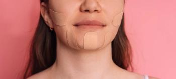
Face taping promises smoother skin and fewer wrinkles without injectables, but evidence suggests any visible benefits are largely temporary.
Cold atmospheric plasma eases stubborn oral lichen planus in long follow-up study, with a new scoring system tracking pain, function, and healing.
Management of oral lichen planus (OLP), a chronic, T cell–mediated inflammatory condition of the oral mucosa, remains challenging, particularly in refractory cases, where standard therapies such as topical or systemic corticosteroids and other immunosuppressive agents may be ineffective or limited by adverse effects. In this context, localized cold atmospheric plasma (CAP) has emerged as a potential alternative, thanks to its anti-inflammatory, antimicrobial, and tissue-regenerative properties.1 A new retrospective case series provides long-term evidence for CAP usage, while also introducing a novel clinical assessment tool designed to better capture disease severity and patient burden.2
The study evaluated 4 patients with histologically confirmed, treatment-resistant OLP, including both erosive and reticular subtypes. All patients underwent intraoral CAP therapy, at a minimum once a week, using an argon-based plasma device between 2016 and 2019, with follow-up extending up to 9 years. Notably, no patient received concurrent immunosuppressive or anti-inflammatory therapy during the study period.
Clinical outcomes were measured using a newly developed Lichen Planus Severity Score (LPSS), which integrates both clinician-assessed parameters (inflammation and ulceration) along with patient-reported outcomes like pain and functional impairment. Each domain is scored on a standardized severity scale, ranging from Grade I (minimal disease) to Grade IV (severe disease). The LPSS was designed to address limitations of existing scoring systems like the Thongprasom, Reticular-Erosive-Ulcerative (REU), Escudier, Oral Disease Severity Score (ODSS), and Oral Mucositis Assessment Scale (OMAS).
“These instruments primarily assess lesion morphology and macroscopic extent, providing limited insight into the underlying inflammatory activity and tissue ulceration that are closely associated with malignant transformation risk, while also neglecting patient-reported outcomes and quality-of-life measures,” the authors wrote.
Across all 4 patients, CAP therapy was associated with substantial and sustained clinical improvement. Baseline LPSS scores ranged from Grade II to Grade III, indicating mild to moderate disease. Following treatment, all patients achieved Grade I status, reflecting minimal or no disease activity. Statistical analysis demonstrated significant negative correlations between treatment duration and all LPSS domains, including inflammation, ulceration, symptoms, and functional limitation, with correlation coefficients ranging from –0.34 to –0.96 (p < 0.001).
Symptomatic improvement, including reductions in pain and functional impairment, typically occurred earlier than visible mucosal healing. On average, patients reported symptom relief after approximately 4 treatment sessions, while macroscopic improvements such as reduced inflammation and ulceration were observed after approximately 6 sessions. A transient increase in inflammatory signs was noted early in treatment before sustained improvement ensued.
Long-term follow-up findings were also notable. Patients maintained clinical remission for years after treatment, with no evidence of malignant transformation in any case. In 1 patient, recurrence occurred at a different anatomical site, while previously treated mucosa remained unaffected. Another patient experienced delayed improvement, likely related to ongoing exposure to dietary irritants, but ultimately achieved sustained disease control.
CAP therapy was well tolerated across all cases. No systemic adverse events were reported during treatment or follow-up. Minor localized effects were limited, with 1 patient reporting transient dentinal hypersensitivity when the plasma device contacted exposed tooth surfaces. Importantly, no evidence of treatment-related complications or carcinogenic effects was observed over the extended follow-up period, which represents the longest reported observation for intraoral CAP therapy to date.
The study also demonstrated that the LPSS is a practical and reproducible tool for monitoring OLP. Interobserver reliability for clinician-assessed parameters was substantial to near-perfect, supporting the consistency of the scoring system. LPSS scores declined consistently with treatment, indicating responsiveness to clinical change and potential utility in both clinical practice and research settings.
Despite these promising findings, the study is limited by its small sample size, retrospective design, and lack of a control group, which restricts conclusions regarding comparative efficacy. Additionally, anatomical limitations of current plasma devices may restrict the treatment of deeper oropharyngeal lesions.
Overall, this study suggests that CAP therapy may offer a safe and effective non-immunosuppressive option for patients with refractory OLP, with durable benefits observed over long-term follow-up. Additionally, the introduction of the LPSS provides a more comprehensive framework for assessing disease severity and treatment response. Larger, controlled studies are needed to validate these findings and further define the potential role of CAP in OLP management.
References
1. Raissi-Dehkordi N, Raissi-Dehkordi N, Ebrahimibagha H, et al. Advancing chronic and acute wound healing with cold atmospheric plasma: cellular and molecular mechanisms, benefits, risks, and future directions. Front Med (Lausanne). 2025;12:1527736. Published 2025 Feb 26. doi:10.3389/fmed.2025.1527736
2. Seebauer C, Metelmann HR, Falougy M, Kaduk W, Kremer L, Daeschlein G. Cold plasma treatment for oral lichen planus: a retrospective case collection and proposal of a new clinical score. J Dtsch Dermatol Ges. Published online April 29, 2026. doi:10.1111/ddg.70193