

News|Articles|July 24, 2026
Masterclass Insights: Optimizing Care for Ulcerated Hemangiomas and Café-au-Lait Macules
Author(s)Marie Bosslett, Assistant Editor
Listen
0:00 / 0:00
Key Takeaways
- High-risk hemangioma ulceration sites include diaper area, lips, neck folds, and intertriginous skin, warranting close monitoring for secondary infection, bleeding, and functional impairment.
- Propranolol accelerates healing and symptom control beyond wound care alone; adjunctive topical timolol, pulsed dye laser, or surgery can be selected by lesion size, location, and response.
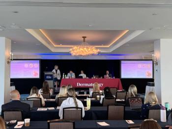
Sarah Chamlin, MD, and Jonathan Dyer, MD, shared practical guidance on treating ulcerated infantile hemangiomas and assessing café-au-lait spots, emphasizing pain control, propranolol, follow-up, and genetics.
Advertisement
At this year’s annual meeting, the
Ulcerated Infantile Hemangiomas
Sarah Chamlin, MD, attending dermatologist at the Ann & Robert H. Lurie Children’s Hospital of Chicago and professor of pediatrics and dermatology at the Northwestern University Feinberg School of Medicine, opened by reviewing ulceration, the most common complication of infantile hemangiomas, noting that ulceration is associated with significant pain, infection risk, bleeding, and permanent scarring. She emphasized that lesions in high-friction or moisture-prone locations—including the diaper area, lips, neck folds, and intertriginous regions—are particularly susceptible and require close monitoring. Families should receive clear instructions regarding gentle cleansing, appropriate dressings, and strategies to minimize friction and secondary infection. Pain management is essential because discomfort often interferes with feeding, sleeping, and routine infant care.
Chamlin reviewed evidence supporting oral propranolol as first-line therapy for ulcerated infantile hemangiomas, highlighting its ability to accelerate healing while simultaneously treating the underlying vascular lesion. Although wound care alone may ultimately lead to healing, propranolol substantially shortens recovery time and improves symptoms in many patients. She also discussed situations in which topical timolol, pulsed dye laser, or surgical intervention may play adjunctive roles, depending on lesion size, location, and response to medical therapy.
Particular attention was given to multidisciplinary management for complex lesions. Ulcerated hemangiomas involving the perineum, airway, or large segmental distributions may warrant evaluation for associated syndromes or consultation with additional specialties. Throughout the discussion, Chamlin stressed setting realistic expectations with families regarding healing time and the likelihood of residual skin changes even after successful treatment.
Café-au-lait Macules
The next part of the masterclass shifted to pigmentary lesions, with Jonathan Dyer, MD, examining one of the most common consultation questions in pediatric dermatology: determining when multiple CALMs warrant concern for an underlying genetic syndrome. Dyer, professor and chair of the department of dermatology at the University of Missouri, Columbia, began by reviewing the clinical characteristics of CALMs, describing them as well-circumscribed, uniformly pigmented tan-to-brown macules that are usually present at birth or develop during early childhood. While a single lesion or a small number of CALMs is common in otherwise healthy children, multiple lesions warrant additional evaluation. He reminded attendees that 6 or more CALMs larger than 5 mm in prepubertal children or greater than 15 mm after puberty remain one of the diagnostic criteria for neurofibromatosis type 1 (NF1), though this criterion should always be interpreted alongside other clinical findings.
The talk emphasized that CALMs are not synonymous with NF1. Dyer reviewed a broad differential diagnosis that includes Legius syndrome, McCune-Albright syndrome, Noonan syndrome with multiple lentigines, constitutional mismatch repair deficiency, Fanconi anemia, and other mosaic or pigmentary disorders. Lesion morphology, distribution, associated pigmentary findings, and the presence of extracutaneous manifestations can all help narrow the diagnosis. Dyer encouraged dermatologists to avoid making premature diagnoses based solely on pigmentary findings in infancy. Instead, he advocated for longitudinal follow-up with serial skin examinations, family history review, developmental assessments, and referrals to genetics when clinical suspicion remains high.
He also highlighted the growing role of molecular testing in children with multiple café-au-lait macules who do not yet meet full clinical criteria for NF1. Genetic testing can help distinguish NF1 from overlapping conditions such as Legius syndrome, allowing clinicians to provide more accurate counseling, surveillance recommendations, and prognostic information while avoiding unnecessary imaging or interventions.
Dyer concluded by emphasizing that the dermatologist often serves as the first clinician to recognize these lesions and initiate an appropriate diagnostic evaluation. Careful documentation, thoughtful longitudinal monitoring, and collaboration with genetics and other pediatric specialists can facilitate earlier diagnosis of associated syndromes while reassuring families when lesions ultimately prove to be isolated, benign findings.
Dermatology Times is on-site in Minneapolis covering SPD’s latest updates from July 22 to 25th; view all our live conference coverage
Advertisement
Related Content
Advertisement



Latest CME


Advertisement
Advertisement
Trending on Dermatology Times
1
How Emerging Androgenetic Alopecia Therapies Target DHT at the Scalp Level
2
Social Media Mythbusters: Beef Tallow
3
Dermatology Times Inaugural NP/PA Connect Cutaneous Oncology Workshop Kicks Off in Tampa
4
25 Years Later: Remembering September 11 Through the Lens of Dermatologic and Burn Care
5