
News|Articles|June 24, 2026
Vitiligo-Like Depigmentation Reported in NHEJ1-Related Radiosensitive SCID
Fact checked by: Yasmeen Qahwash
Listen
0:00 / 0:00
Key Takeaways
- NHEJ1-related SCID typically manifests as TB−NK+ immunodeficiency with microcephaly, growth failure, and radiosensitivity, but pigmentary abnormalities have not been considered part of its canonical presentation.
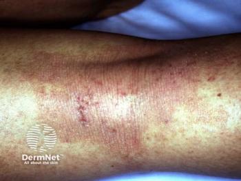
- Cutaneous findings included ~90% body surface depigmentation, early-onset dermatitis, and mixed black/silvery-white hair, with systemic infections and immune cytopenias prompting whole-exome confirmation.
A recent JAAD case report describes congenital depigmentation and progressive posttransplant repigmentation in a child with NHEJ1-related radiosensitive SCID—a pairing not previously documented.
Advertisement
A new case report published in
NHEJ1-related SCID arises from pathogenic variants in the gene encoding Cernunnos-XLF, a scaffolding protein required for DNA end ligation during nonhomologous end-joining repair. The disorder typically presents with a TB-NK+ immunophenotype, microcephaly, growth retardation, and radiation sensitivity. Pigmentary abnormalities have not factored into its recognized clinical spectrum, unlike depigmentation syndromes such as Chediak-Higashi, Griscelli, and Hermansky-Pudlak, which stem from melanosome transport defects rather than DNA repair pathway disruption.1
Case Findings and Posttransplant Repigmentation Pattern
The patient was born at term to biologic parents with no family history of vitiligo. Her skin appeared lighter than expected from birth, and she developed a generalized pruritic erythematous rash within the first month of life. Complete hair loss occurred at 3 months of age. At 5 months, dermatologic examination revealed well-demarcated depigmented patches covering approximately 90% of body surface area, without palmoplantar keratoderma, nail dystrophy, or dental abnormalities, with scalp hair described as sparse, fine, and black.1
Her medical history included oral thrush, pneumonia, recurrent otitis media, and failure to thrive, with laboratory findings of lymphopenia and hypogammaglobulinemia. Whole exome sequencing in May 2023 confirmed a homozygous pathogenic NHEJ1 variant. At 9 months, she underwent matched-sibling HSCT without conditioning chemotherapy due to radiosensitivity risk, with an uncomplicated posttransplant course and no clinical signs of acute or chronic graft-vs-host disease (GVHD).1
By 2 to 3 months post transplant, faint tan macules appeared on the trunk and extremities, coalescing into areas of repigmentation. One year later, the patient showed diffuse residual depigmentation alongside sharply defined islands of normal pigmentation, most prominent on the face, hands, and feet, with scalp hair regrown as a mix of black and silvery-white strands. Repigmentation continued through 2025, though it remained incomplete at last follow-up.1
Proposed Mechanisms Behind the Pigmentary Changes
The authors offer 2 nonexclusive explanations for the depigmentation. The first centers on immune dysregulation: Aberrantly developing T or B cells in SCID may become autoreactive and target melanocytes, a mechanism described in related DNA repair disorders including Artemis and LIG4 deficiency, which share the NHEJ1 repair pathway. The second points to intrinsic melanocyte fragility, reasoning melanocytes lacking functional DNA repair may be more vulnerable to ultraviolet and oxidative damage.1
The authors considered GVHD as an alternative explanation, as maternal T-cell engraftment occurs in up to 40% of patients with SCID and can produce GVHD-like skin findings, sometimes prior to transplantation itself. They favored a vitiligo-like mechanism instead, citing the absence of other clinical GVHD features and a repigmentation pattern tracking with immune reconstitution rather than following a fixed or inflammatory course. Histopathologic confirmation of vitiligo was not obtained.1
Alhaider and colleagues noted repigmentation after HSCT has been reported previously in patients with SCID broadly but not specifically in those with NHEJ1 mutations, citing a
The authors suggest recognizing this association may help clinicians distinguish autoimmune pigmentary signs from GVHD in immunodeficient patients and anticipate the potential for recovery after transplantation. They identify this as the first reported case linking pigmentary change with NHEJ1 deficiency, raising the possibility that immune or DNA repair mechanisms contribute to melanocyte homeostasis more broadly.1
References
- Alhaider A, Almutairi M, Alomari A, Altuwaijri A, Almarshoud G, Alakrash L. Vitiligo like depigmentation in a patient with NHEJ1 deficiency related radiosensitive SCID: a case report. JAAD Case Rep. 2026;73:106-110. doi:10.1016/j.jdcr.2026.05.010
- Heath CR, Burk CJ, Lawley LP, Mancini AJ, Connelly EA. Severe combined immunodeficiency (SCID)-associated dyschromia with subsequent repigmentation: a report of two patients. Pediatr Dermatol. 2009;26(2):162-8. doi:10.1111/j.1525-1470.2009.00876.x
Advertisement
Related Content
Advertisement


Latest CME


Advertisement
Advertisement
Trending on Dermatology Times
1
Our Current Gap In Melanoma Detection
2
The New Economics of Dermatology Claims: Understanding and Responding to Rising Denials
3
Striking the Balance with JAK Inhibitors in Atopic Dermatitis Management
4
Vitiligo-Like Depigmentation Reported in NHEJ1-Related Radiosensitive SCID
5