Patient Presentation of PsA and PsO
Drs Mark G. Lebwohl and Joseph F. Merola discuss how patient presentation differs in patients with PsO and PsA.
Episodes in this series

Mark G. Lebwohl, MD: Let’s move on to the next question. Joe, how does a patient with psoriasis typically present? How does the presentation change when a patient progresses to psoriatic arthritis [PsA]?
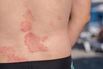
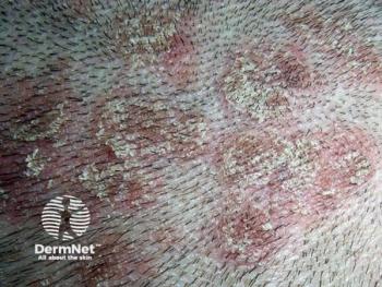
Joseph F. Merola, MD, MMSc: That’s a very broad question. This is a good point for us to talk a about what some would call domains of disease. In this context, let’s try to frame the different phenotypes that might present. We’re talking mostly about patients with plaque psoriasis and scalp psoriasis. The audience is quite familiar with that presentation. I mentioned already some of the at-risk phenotypes in that population—nail disease, scalp disease, inverse or intertriginous psoriasis—all of which we believe increase or at least are associated seemingly with an increased risk of developing psoriatic arthritis. It’s interesting to talk a little about how that patient presents with PsA in this context. That’s also varied, but let me start with the possible domains of disease.
We have patients who present with peripheral arthritis. That in itself can come in multiple flavors. We’ll come back to that. There are patients who can present with axial psoriatic arthritis or spine involvement. That involves sacroiliitis but may also present with axial or spine involvement anywhere along the axial skeleton. That’s important. Those patients also may more commonly present with certain joints, like hip involvement and others that cluster with that phenotype. They may come more with dactylitis with plantar fasciitis and other clinical phenotypes. We have patients who may present predominantly with enthesitis. That’s inflammation at sites that have tendon insertion into bone. We could talk more about that, but certain classical areas—epicondylitis at the elbow, an Achilles’ insertion, enthesitis, or plantar fasciitis—are more common sites that might present.
We have multiple subphenotypes around there. There’s dactylitis or the so-called sausage digit that might present as well. If a patient presents with an entire digit that’s swollen, not just the peripheral joint itself but the intervening soft tissue is inflamed, that’s yet another phenotype. What’s interesting is these are not mutually exclusive, so it turns out that any given patient over time can morph or change and develop these different manifestations over their illness. For us in dermatology, it’s more obvious.
Mark knows this very well from his practice: patients who presents with a red, hot, swollen knee or a big, fat swollen digit are going to be more obvious in their presentation and may warrant a discussion with the rheumatologist. That depends on your diagnostic and management comfort, but I’ll talk about that achy patient here. The patient who presents with psoriasis in the dermatology clinic, who has joint complaints; they’re achy. They haven’t put a finger on what it is. They have some stiffness. Maybe they didn’t tie their recurrent tendonitis into this larger presentation of psoriatic arthritis. Perhaps they’ve had recurrent plantar fasciitis, they have some nonspecific low-back pain. Those are the patients for whom we’re going to have to tease out is this psoriatic arthritis. We may be the ones making that connection between skin disease and muscular skeletal complaints, whether they’re presenting early or haven’t made that connection.
Mark, what’s your experience with the presentation, particularly in the dermatology office, in front of the dermatologist with that patient who’s presenting with early symptoms of PsA?
Mark G. Lebwohl, MD: I like to put numbers on these. I love the way you divided it into the different forms of psoriasis. Ultimately, plaque psoriasis is the predominant presenting form of psoriasis, although close to 20% can come in with guttate psoriasis. Very rare are postular erythrodermic, but those are the deadly ones. Of course, in plaque, we include inverse. Palm and sole psoriasis is separate, less common than plaque psoriasis.
In terms of the arthritis presentation that you asked about, there are several forms. When you see dactylitis, that patient has psoriatic arthritis. I’ve made that diagnosis based on only dactylitis. The patient had no evidence of psoriasis anywhere but responded dramatically to the treatments that we know help psoriatic arthritis, TNF [tumor necrosis factor] blockers, or IL-17 blockers. I’ve had that happen twice in my career—individuals came with just dactylitis.
Joseph F. Merola, MD, MMSc: I have to pause there because I was excited to hear you say that. I just came from the clinic for this call. One of my patients in the middle of the clinic was a gentleman who presented with plaque psoriasis. We called in all the residents to look at this. Dactylitis of his second digit. To your point, it’s so specific to this spondylitis camp that I felt confident saying to this guy, “We know you have plaque psoriasis, but you have psoriatic arthritis.” This is not 1 of those soft calls where I have to say, “Let’s get some films. Let’s get some inflammatory markers.” We got the diagnosis. To your point, it’s a great feeling to tell a patient that.
Mark G. Lebwohl, MD: Exactly. Because you just said that, when a patient comes to me with back pain, I know it’s not me because my friends who are rheumatologists see the same patient and don’t know. Back pain is so common.
Joseph F. Merola, MD, MMSc: That 1 is more the discussion of the hair loss patient. Even we wince a little bit at the back, but we have to dig into it. We have to dive into it, but yes, much less clear for sure.
Mark G. Lebwohl, MD: All of us see those patients and with the drugs available, they’re so effective and safe that I even use them as a diagnostic tool. If they respond to them, we know it was psoriatic arthritis, not osteoarthritis. Those are very challenging, but putting them into large categories, most patients we see have 1 or a few peripheral joints involved, so it’s monoarthritis or oligoarthritis. They respond well. They can be very destructive even though it’s 1 or a few joints, but they respond very well to some of the new treatments we have.
I’ll put TNF blockers, IL17 blockers, and JAK inhibitors in the category of treatments great for psoriatic arthritis. More recently, some of the IL-23 blockers have shown some decent evidence that they’re helpful in psoriatic arthritis. Somebody has 1 or a few peripheral finger joints. It’s an easier diagnosis when they have signs of psoriasis. When they don’t have signs of psoriasis, the rheumatologist is scratching his head saying, “It could be psoriatic arthritis but I’m not sure.” Then they send him to me and say, “Can you find psoriasis in that patient anyplace?” Because they want to justify using these drugs that we have that are pretty good.
Joseph F. Merola, MD, MMSc: That’s a great segue. We’re going to come back to the case later on but also several other more nuanced topics. I love your description of the MOAs [mechanisms of action] that face this disease quite well. At the same time, there’s nuance. We’ll cover that, in terms of phenotypic subsets of disease and if they’re damaging joints. Is there what we would call radiographic progression on the ground in terms of the data pieces? Do they have actual diseases? That’s going to be potentially a slicing-dicing moment in which we think about which treatment class we might we turn to, and where we have more and less data. I also love that you brought up even the nuance of peripheral arthritis. Is it monoarticular, oligoarticular, or polyarticular arthritis? So much of the data we look is based on polyarticular arthritis, but that probably extrapolates very well into these other subtypes, including oligoarticular and even monoarticular. All points are well taken as we unpack this.
Mark G. Lebwohl, MD: The difference when they have polyarthritis is rheumatoid arthritis. The real difference in the rheumatoid factor is either it’s nonexistent, or it’s low and the patient has psoriasis. That’s how you distinguish those 2. They can get the same exact defects—the swan-neck deformity, the boutonniere deformity—that you see in rheumatoid arthritis. But the rheumatoid factor is negative, and the psoriasis is there. That’s the other presentation.
Joseph F. Merola, MD, MMSc: We won’t scare anyone off. Probably 8% to 12% of patients have a low tighter RF [radiofrequency] or CCP [cyclic citrullinated peptide]. I don’t worry about those patients. Of course, Mark’s referring to those higher, tighter ones that would be very focused on RA [rheumatoid arthritis]. I’ll tell you, for the head-scratcher, that we have some patients who we absolutely swear have psoriasis and RA. They have a high tighter disease. They have x-rays that look like RA and lucky for us, many of our therapies, including anti-TNFs and others, treat both. It works out quite well in those scenarios.
Transcript edited for clarity
Newsletter
Like what you’re reading? Subscribe to Dermatology Times for weekly updates on therapies, innovations, and real-world practice tips.


