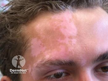
Initiating Patient Conversations Around Vitiligo Management
Experts in dermatology share their approaches to communicating with patients about their vitiligo, highlighting family history and genetic awareness.
Episodes in this series

Nada Elbuluk, MD: Do you get into the discussion about genetic risk with patients? That can be a big concern for some people.
David Rosmarin, MD: Yes, people in their 30s or even 20s who are thinking about children. They often want to know, “Will I pass this on to my child? Did I get this from 1 of my parents?” Or “I think this relative had it.” About 1% of the population, 0.5% to 2% in some estimates, will have vitiligo. If you have a first-degree relative, then your risk is increased about 4%. It’s about 6-fold higher. It’s complex, and there’s a genetic component, a predisposition. But chances are, a parent will not have a child with vitiligo. That can be reassuring to patients. They also want to know if they can do screening tests to determine that. Unfortunately, we don’t have the capacity to know in advance if a child will have it.
Nada Elbuluk, MD: I get a lot of patients who will say, “I don’t have anyone in my family who has vitiligo.” I explain that there are other autoimmune diseases that are correlated. Many times, when I’ll start asking, “Does anyone in your family have thyroid disease, type 1 diabetes, or anything else?” A high percentage of people have some extended family member who has autoimmune diseases. It’s not a direct linear correlation, which sometimes makes it harder for people to understand. As you said, it’s complex. Genetics is part of it, and other autoimmune diseases are also part of it. Trying to break that down for patients is important.
David Rosmarin, MD: Besides screening, when you’re doing a physical exam on a patient, do you always use a Wood’s lamp? Sometimes? How do you use the Wood’s lamp?
Nada Elbuluk, MD: Good question. I always have a Wood’s lamp in my patient room whenever I’m seeing people with vitiligo or other pigmentary disorders. With a darker-skinned patient, it’s very clear. You don’t always need a Wood’s lamp. You can see the areas that are depigmented. I use the Wood’s lamp in the majority of cases. Particularly for individuals with lighter- to medium-toned skin, there’s a lot more that you can see with the Wood’s lamp, that you don’t always see with the naked eye. The Wood’s lamp is important. I always like my patients to be in a gown because I want to see their total percentage of body surface area. Sometimes they’ll come in and say, “I just care about what’s on my face.” I still want to see what percentage is involved. That way, when I’m advising them about treatment, I can give a range of options, even if they’re only concerned about 1 area. What’s your approach?
David Rosmarin, MD: It’s helpful to look everywhere. The Wood’s lamp is as an essential tool, especially for those who are fair-skinned. It’s not infrequent that a patient comes in saying that they have limited disease, but then you look at it on a Wood’s lamp and realize it’s more extensive. That can be an emotional shock to a patient, when they’re looking at their skin under the Wood’s lamp. They say, “Wow, I didn’t realize how extensive it is.” That’s an emotional moment, and they need our support.
Nada Elbuluk, MD: Absolutely.
David Rosmarin, MD: It’s an essential tool. I’m always prepared with tissues at my bedside if needed.
Nada Elbuluk, MD: I do the same thing. I have a tissue box in every room because it can be emotional for many people. How do you like to approach the conversation with treatment, and your recommendations when patients come in?
David Rosmarin, MD: When I see a patient, the first thing I try to do is group them into 1 of 2 categories. Is it progressive or stable disease? They may tell you that it’s progressing and have pictures to back that up. But there may also be physical exam findings that indicate this is progressive disease, such as confetti regions, which are 1- to 2-mm white macules. They may have trichrome vitiligo, or 3-colored vitiligo, with normal skin, depigmented skin, and a color in between. They may have Koebner phenomenon, with a scratch on their body indicating new onset of vitiligo. The fourth, which is very uncommon, is inflammatory vitiligo. It’s a red rim around the vitiligo lesions. Not redness throughout, but around the rim of the vitiligo. But that’s very uncommon.
If I see that and realize this disease is progressing, I try to halt the progression with dexamethasone, from 2 to 5 mg, on 2 consecutive days of the week for about 12 weeks. The data are fairly good about how that can halt the progression in more than 90% of patients. If not, you can always go for longer, or increase the dose of oral steroids. I’ll also offer phototherapy on that initial visit, as a standard way to try to halt progression and start the repigmentation process. But in my experience, a minority of patients, especially those that come from far, end up doing phototherapy because it’s burdensome to miss school or to miss work to do it. Patients don’t realize how many sessions you need, to give it a fair shot. Usually, it’s about 48 sessions in the guidelines. But some papers indicate that some patients need over 72 sessions before they respond. The key point is vitiligo takes time, no matter what modality.
Transcript edited for clarity
Newsletter
Like what you’re reading? Subscribe to Dermatology Times for weekly updates on therapies, innovations, and real-world practice tips.


