

News|Articles|May 13, 2026
Beyond Breslow Depth: Using GEP to Inform SLNB Decisions in T1 and T2 Melanomas
Author(s)Marie Bosslett, Assistant Editor
Listen
0:00 / 0:00
Key Takeaways
- GEP assays are being adopted as adjuncts to Breslow depth and ulceration to individualize SLNB decisions in select T1b–T2a patients with uncertain nodal risk.
- A low-risk i31-GEP (Class 1A) aligning with stage IA features can support omission of SLNB and imaging, with many clinicians using RFS >90% as a low-risk benchmark.
Clinicians discuss how melanoma gene expression profiling guides sentinel node biopsy choices, refines early-stage risk, and shapes personalized surveillance and oncology referral.
Advertisement
Dermatology clinicians from across Florida convened for a
Although participants varied in how often they order GEP tests, the group agreed that these assays are becoming a valuable adjunct to standard staging, particularly for T1 and T2 melanomas in the “gray zone” where SLNB is discretionary.
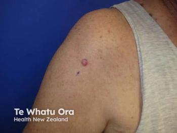
Case #1: When “Low Risk” Staging Feels Incomplete
The first case involved a woman aged 45 years with a plantar melanoma (Breslow depth 0.6 mm, nonulcerated, mitotic rate 1, no lymphovascular invasion or regression), staged as pT1a/stage IA. Under current National Comprehensive Cancer Network (NCCN) guidance, she would not routinely qualify for SLNB. Most panelists agreed that wide local excision alone would be appropriate on the basis of standard staging. However, several raised concerns specific to acral melanoma biology and the known limitations of current staging systems in thin disease.
“This is actually where I think GEP testing really does come into play and shine,” one attendee said. “[This] is how I use it and consider it clinically.”
In this case, an i31‑GEP test was obtained and returned a low‑risk (Class 1A) result, with:
- Predicted SLNB positivity: 4.7%
- Recurrence‑free survival (RFS): 91.3%
- Distant metastasis‑free survival: 94.6%
- Melanoma‑specific survival: 97.8%
These data were concordant with her favorable stage IA status. Moody and the participants confirmed that this result supported proceeding with wide local excision and dermatologic follow‑up, without SLNB or additional imaging. In several practices, an RFS rate greater than 90% is considered a “low‑risk” threshold that justifies standard surveillance alone.
Case #2: Using GEP to Clarify the SLNB “Discuss and Consider” Zone
The second case featured a man aged 63 years with a back melanoma (0.8 mm, nonulcerated, mitotic rate 2), staged as pT1b and therefore falling into the NCCN “discuss and consider” group for SLNB. Many clinicians view this as one of the most challenging decision points in early melanoma care. Polling among the attendees showed that risk-benefit of the procedure and GEP results were the most influential factors guiding SLNB recommendations, more so than pure access to oncology or surgery.
Moody’s discussion referenced the MERLIN_001 trial (
In this case, a clinicopathologic GEP test was obtained and returned a low‑risk result. The patient—already hesitant about surgery due to caregiving responsibilities for his wife—ultimately declined SLNB and underwent wide local excision alone, followed by dermatologic and nodal exams every 3 months.
Case #3: When GEP and Staging Diverge
A third case highlighted how GEP can reveal higher biological risk in what appears, on paper, to be early disease. An outdoor worker aged 58 years presented with a forearm melanoma (1.0 mm, nonulcerated, mitotic rate 1) staged as pT1b. Most panelists favored SLNB and GEP testing. The i31‑GEP returned a high‑risk (Class 2B) result, with predicted SLNB positivity of 13.1% and RFS rate of 84%.
Here, GEP and traditional staging were discordant: clinically borderline, but molecularly high risk. About 40% of attendees said they would prioritize the GEP result if clinical features align, and the majority would involve a multidisciplinary team of surgical and medical oncologists in decision‑making. One attendee noted that although adjuvant systemic therapy for stage I disease is not standard, GEP‑high patients are “not typical stage I” biologically, and closer monitoring is warranted.
“I think the only thing you can really do is stress the importance of close clinical follow‑up...I would definitely involve a medical oncologist,” another attendee added.
The group consensus was to:
- Proceed with SLNB
- Treat a negative SLNB but high‑risk GEP as a trigger for enhanced surveillance, including more frequent skin and nodal exams and, in many practices, baseline and/or periodic imaging
- Involve medical oncology early, even for stage I patients with a high‑risk molecular profile
Final Thoughts
Overall, the roundtable underscored that although Breslow depth and ulceration remain foundational, GEP testing is increasingly used as a practical, real‑world tool to personalize risk assessment, SLNB decisions, and follow‑up intensity for early melanoma. Most panelists already use at least one commercially available GEP assay. However, they identified clearer NCCN and American Academy of Dermatology guidance and more formal algorithms as the changes that would most increase their confidence and standardize use. Tumor boards and medical oncologists also remain central to translating GEP results into long‑term management plans.
Advertisement
Related Content
Advertisement


Latest CME


Advertisement
Advertisement