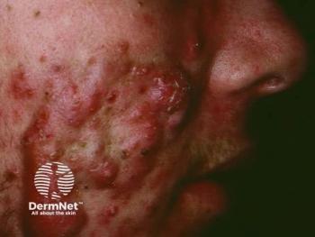
Persistent acne sequelae, including post-inflammatory erythema (PIE), post-inflammatory hyperpigmentation (PIH), and atrophic scarring, showed photographic improvement following individualized protocols combining photothermal biomodulated autologous platelet-rich plasma (PTBM-PRP), laser therapy, and light-emitting diode (LED) phototherapy in a recent 3-patient case series.1
Published in Case Reports in Dermatology, the retrospective report evaluated 3 women with distinct acne-related concerns using serial photographs obtained during routine clinical follow-up. Because validated acne severity, pigmentation, erythema, and scar grading scales were not applied prospectively, investigators conducted post hoc semi-quantitative assessments where image quality permitted. Follow-up ranged from 1 month to 1 year.1
The authors emphasized that the report was designed to describe the feasibility, tolerability, and photographic evolution of the treatment approach rather than establish efficacy or determine the contribution of individual treatment modalities.1
Individualized Protocols Target Different Acne Sequelae
Each treatment protocol incorporated autologous PRP that underwent photothermal biomodulation before administration. PRP was processed using the MCT Unit (Meta Cell Technology) with an exosome-targeted device setting of 467 nm, 1 J/cm², and 37°C for 10 minutes. Treatment delivery and accompanying device-based therapies were then tailored to each patient's primary clinical concern.1
The first case involved a 21-year-old woman with grade II inflammatory acne and persistent PIE. She underwent 3 treatment sessions at 3-week intervals followed by a maintenance session at 6 months. PTBM-PRP was administered intradermally, followed by microneedling and multi-wavelength LED phototherapy incorporating blue, red, yellow, and near-infrared wavelengths.1
Frequently Asked Questions
What is PTBM-PRP?
PTBM-PRP refers to autologous platelet-rich plasma that undergoes photothermal biomodulation before administration. In this case series, investigators used an exosome-targeted device setting before administering the PRP as part of individualized multimodal treatment protocols.1
What acne sequelae were treated?
The 3 cases involved PIE with active inflammatory acne, PIH with residual inflammatory acne, and atrophic boxcar and rolling acne scars.1
Did the study prove that PTBM-PRP improves acne sequelae?
No. The uncontrolled 3-patient case series documented photographic improvement but was not designed to establish efficacy or causality. Because patients received multiple therapies, the individual effect of PTBM-PRP also could not be determined.1
Retrospective photographic assessment estimated approximately 35 to 45 visible inflammatory lesions at baseline compared with approximately 8 to 12 at 6 months. In the available 1-year photographic field, 0 to 3 residual inflammatory lesions were visible. PIE decreased from severe at baseline to mild at 1 year. Overall photographic improvement was estimated at approximately 60% to 70% at 6 months and 80% to 90% in the available 1-year field, although the authors cautioned that differences in field of view and nonstandardized photographic conditions limit interpretation.1
PIH and Residual Acne Show Changes at 1 Month
The second case involved a 32-year-old woman with PIH, uneven skin tone, and residual inflammatory acne. She received a single Fotona 4D laser session using neodymium-doped yttrium aluminum garnet (Nd:YAG; 1064 nm) and erbium-doped yttrium aluminum garnet (Er:YAG; 2940 nm) wavelengths, followed by PTBM-PRP delivered with microneedling and multi-wavelength LED phototherapy.1
At 1 month, visible inflammatory lesions were estimated to have declined from approximately 8 to 12 at baseline to 3 to 5. PIH and skin tone unevenness improved from moderate-to-severe to mild-to-moderate, with an overall photographic improvement estimated at approximately 40% to 60%. The authors noted that the short follow-up and differences in lighting may have influenced the apparent improvement in pigmentation.1
Atrophic Acne Scarring Improves at 6-Month Follow-Up
In the third case, a 26-year-old woman with shallow boxcar and rolling atrophic acne scars underwent a single fractional ablative Er:YAG laser session followed by PTBM-PRP administered using the nappage technique and LED phototherapy. At 6 months, retrospective photographic review suggested a moderate reduction in scar visibility, shadowing, and texture irregularity, with an estimated photographic improvement of approximately 40% to 50%.1
The before-and-after images on pages 5, 7, and 8 illustrate the photographic changes reported for the 3 patients, but they also show why the authors' caveats about differences in lighting, angle, and field of view are important when interpreting the findings.
Safety and Biological Rationale for PTBM-PRP
Treatment was generally well tolerated across the 3 cases. Reported adverse effects were limited to transient local reactions, including erythema and mild edema, which resolved spontaneously. No serious adverse events were reported.1
The rationale for PTBM-PRP centers on the possibility that photothermal or photobiomodulatory conditions may influence platelet-derived signaling and extracellular vesicle release. However, the authors stressed that no exosome quantification, platelet activation marker assessment, or mechanistic biomarker analysis was performed in these patients. Therefore, the biological effect of photothermal biomodulation in the reported cases remains hypothetical.1
Previous research has suggested that PRP used alone or alongside energy-based devices may support collagen remodeling and improve outcomes in acne scarring. Fractional laser therapy similarly promotes dermal remodeling through controlled injury, while LED phototherapy has been studied for anti-inflammatory, antimicrobial, and regenerative effects depending on the wavelengths used.1,3
Study Limitations and Need for Controlled Trials
Importantly, the multimodal design means the observed changes cannot be attributed specifically to PTBM-PRP. All patients received combination treatment involving PTBM-PRP and laser and/or LED therapy, preventing investigators from determining the contribution of each modality or establishing whether their effects were additive or synergistic.1
Additional limitations included the sample size of only 3 patients, absence of a control group, randomization, or blinded assessment, lack of prospectively validated outcome measures, nonstandardized photography, differing follow-up periods, and absence of formal patient-reported outcome measures.1
The authors concluded that the cases support the clinical feasibility and tolerability of integrating PTBM-PRP with laser and LED therapy in individualized protocols for acne sequelae but do not establish efficacy or causality. Prospective controlled studies using standardized protocols and objective outcome measures are needed to determine the role of the approach.1
References
- Labidi Menif Y, Cordero L. Photothermal biomodulated autologous PRP combined with laser and LED phototherapy for acne sequelae: a report of three illustrative cases. Case Rep Dermatol. 2026;18:559-569. doi:10.1159/000553563
- Karadag AS, Aslan Kayıran M, Wu CY, Chen W, Parish LC. Antibiotic resistance in acne: changes, consequences and concerns. J Eur Acad Dermatol Venereol. 2021;35(1):73-78. doi:10.1111/jdv.16686
- Min S, Yoon JY, Park SY, Moon J, Kwon HH, Suh DH. Combination of platelet rich plasma in fractional carbon dioxide laser treatment increased clinical efficacy for acne scar by enhancement of collagen production and modulation of laser-induced inflammation. Lasers Surg Med. 2018;50(4):302-310. doi:10.1002/lsm.22776