|Articles|November 7, 2022
- Dermatology Times, February 2023 (Vol. 44. No. 02)
- Volume 44
- Issue 02
An Overview of Argyria
Author(s)Peter Baek , Bernard A. Cohen, MD
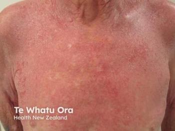
Argyria is a rare dermatologic condition that presents with blue or grey mucocutaneous discoloration following cutaneous exposure or ingestion of silver.
Advertisement
Disease Overview
Argyria is a rare dermatologic condition that presents with blue or grey mucocutaneous discoloration following cutaneous exposure or ingestion of silver. It can present with blue-grey discoloration of the skin, eyes, nails, and gums. Further discoloration can be exacerbated with sunlight exposure. While classified as benign condition, once afflicted, skin discoloration is irreversible. Given the rare nature of argyria, this condition is often a diagnosis of exclusion and can be mistaken for other dermatologic conditions.1
Epidemiology
With the decline of heavy silver exposure and medicinal use, argyria is a rare condition. Given the extreme rare nature of argyria, there is no reported prevalence statistics of argyria. However, through case reports, it has been determined that the most common etiology of argyria is workplace exposure and medicinal use. Given the transmission mechanism of argyria, argyria can affect all individuals regardless of age, sex, gender, and ethnicity. The elemental form of silver is a white transition metal. Among metals, silver has the highest conductivity, thermal conductivity, and reflectivity, which lends to its versatility.2 Occupational silver exposure includes jewelers, silver miners, and silversmiths who have the highest risk of transdermal, transmucosal, or inhaled silver transmission.3 A smaller subset of patients with argyria will be due to medicinal exposure. Its antimicrobial capacity via Ag ion damaging bacterial membranes promoted the use of silvers for medicinal purposes prior to the discovery of more potent antibiotics. Despite the declining use of silver in modern medicine, the FDA still approves a limited subset of products with silver such as Ophthalmic silver nitrate for gonorrheal ophthalmia neonatorum, cutaneous silver sulfadiazine for wounds secondary to second and third-degree burns, and Silver impregnated catheters and endotracheal tubes as an antimicrobial adjunct.4 While medicinal use of silver is declining, there is a niche homeopathic market for colloidal silver as a cure-all therapy that can be obtained online and over-the-counter.4
Diagnosis and Presentation
Due to the rare nature of argyria, it is often a diagnosis of exclusion. If a physician suspects argyria as a potential differential, detailed history such as silver exposure through work or medicinal exposure are noted. The physical exam will often involve an area of the dermis with blue-grey discoloration. Dermatologists also incorporate the use of dermatoscopes to observe for histological patterns such as the silvery papillary dermis to aid in narrowing differential diagnoses.5 The gold standard is a skin biopsy in the affected area. Histopathology will display microgranules ranging from brown to black. The deposition often is linear in formation from the basement membrane of eccrine glands.6,7 Granules can also be found in the papillary dermis in elastic and collagen fibers. Microscopy with hematoxylin and eosin stains can be used for confirmation of silver granule deposits.2
Within the broad umbrella of argyria, there are 3 main subtypes, generalized argyria, localized argyria, and argyrosis. Generalized argyria results from systemic exposure to silver and manifests with a blue-grey or diffuse metallic appearance of the dermis. Areas with increased sun exposure has a higher risk of increased grey pigmentation. A subset of generalized argyria is azure lunula, which is blue-grey discoloration of the nail lunula. The second subtype is localized argyria. This condition is due to the localized cutaneous deposition of silver. Clinical manifestation often includes dark macular spots that range from blue-grey to a more intense black hue. A common example of localized argyria is amalgam tattoo, which results from restorative dental procedures. During tooth restoration, there is impregnation of dental silver amalgam into the oral mucosa, causing a dark blue lesion around the surgical site. The third variant of argyria is argyrosis. Argyrosis is primarily silver deposition in the eye within the Descemet membrane. Patients will present with small dark lesions with green and brown hues.5
Argyria Management
Argyria is a condition that causes permanent discoloration and does not have well established effective treatments. There have been experimental attempts such as chelation, dermabrasion, and hydroquinone, but none have reported long-lasting results.8,7
There have been limited novel studies that have reported improvements with the use of laser treatments. There have been 11 case reports document the use of the 1064 nm Nd:Yag laser and 755 nm alexandrite lasers, with varying treatment parameters. Collectively, each case study reported good outcomes for skin discoloration removal. However, a barrier was the severe pain during the laser removal treatment.9 Long-term follow-ups demonstrate the majority of patients do not have pigment recurrence. However, there is one documented case with recurrence of discoloration 1 year after the completion of therapy. 10,11 In order to further establish laser removal as an efficacious and safe treatment for argyria, appropriate measurements for pain control during treatment is a necessary.
References
1. Jerger SE, Parekh U. Argyria. In: StatPearls. StatPearls Publishing; 2022. Accessed October 16, 2022. http://www.ncbi.nlm.nih.gov/books/NBK563123/
2. Dudeja L, Dudeja I, Janakiraman A, Babu M. Ocular argyrosis: A case with silver deposits in cornea and lens. Indian J Ophthalmol. 2019;67(2):267-268. doi:10.4103/ijo.IJO_730_18
3. Beutler BD, Lee RA, Cohen PR. Localized cutaneous argyria: Report of two patients and literature review. Dermatol Online J. 2016;22(11):13030/qt4wm1j7pt.
4. Exposure-Related Health Effects of Silver and Silver Compounds: A Review. Ann Occup Hyg. Published online June 17, 2005. doi:10.1093/annhyg/mei019
5. Mota L, Dinis-Oliveira RJ. Clinical and Forensic Aspects of the Different Subtypes of Argyria. J Clin Med. 2021;10(10):2086. doi:10.3390/jcm10102086
6. Lencastre A, Lobo M, João A. Argyria -- case report. An Bras Dermatol. 2013;88(3):413-416. doi:10.1590/abd1806-4841.20131864
7. Molina-Hernandez AI, Diaz-Gonzalez JM, Saeb-Lima M, Dominguez-Cherit J. Argyria after Silver Nitrate Intake: Case Report and Brief Review of Literature. Indian J Dermatol. 2015;60(5):520. doi:10.4103/0019-5154.164427
8. Bracey NA, Zipursky JS, Juurlink DN. Argyria caused by chronic ingestion of silver. CMAJ Can Med Assoc J J Assoc Medicale Can. 2018;190(5):E139. doi:10.1503/cmaj.171374
9. Almurayshid A, Park S, Oh SH. Effective laser treatment options for argyria: Review of literatures. J Cosmet Dermatol. 2020;19(8):1877-1882. doi:10.1111/jocd.13549
10. Han TY, Chang HS, Lee HK, Son SJ. Successful treatment of argyria using a low-fluence Q-switched 1064-nm Nd:YAG laser. Int J Dermatol. 2011;50(6):751-753. doi:10.1111/j.1365-4632.2010.04796.x
11. Krase JM, Gottesman SP, Goldberg GN. Recurrence of Argyria Post Q-Switched Laser Treatment. Dermatol Surg Off Publ Am Soc Dermatol Surg Al. 2017;43(10):1308-1311. doi:10.1097/DSS.0000000000001104
Articles in this issue
almost 3 years ago
Vitiligo in Different Ethnicitiesalmost 3 years ago
Rosacea and Menopausealmost 3 years ago
Nonsteroidal Treatment Options for Plaque Psoriasisalmost 3 years ago
An Overview of Documented Flares in Patients With GPPalmost 3 years ago
The Art of Smelling Goodalmost 3 years ago
Losing a Malpractice Case May Depend on Where You Livealmost 3 years ago
The Role We Play in Addressing Racial Disparity in Dermatologyabout 3 years ago
Pemphigus: Updated Review and Emerging TherapiesNewsletter
Like what you’re reading? Subscribe to Dermatology Times for weekly updates on therapies, innovations, and real-world practice tips.
Advertisement
Related Content
Advertisement
Latest CME


Advertisement
Advertisement