

|Articles|February 6, 2019
- Dermatology Times, February 2019 (Vol. 40, No. 2)
- Volume 40
- Issue 2
Skin issues that affect patients with skin of color
Author(s)Maria L. Espinosa, B.S., Peter Lio, MD
Healthcare disparities exist in all fields of medicine, including dermatology. We seek to address these disparities in this article by outlining the differences in epidemiology, presentation, access and outcomes of five conditions in patients with skin of color.
Advertisement
Healthcare disparities exist in all fields of medicine, including dermatology. Despite movements to educate the general public, many myths related to dermatology still exist, such as the belief that those with darker skin tones do not need to protect their skin because they have a minimal risk of developing skin cancer. While it is true that increased amounts of melanin and the mechanisms related to melanosomes in people with skin of color do
The purpose of this brief review is to focus on some differences in epidemiology, presentation, access and outcomes in different skin types for five dermatologic conditions: atopic dermatitis, melanoma, hidradenitis suppurativa, melasma, and vitiligo.
ATOPIC DERMATITIS
In terms of presentation, African Americans have increased risk for post-inflammatory dyspigmentation, plaques on extensor surfaces instead of traditional flexor surfaces and have scattered distinct plaques; while Asian patients more likely to have scaling and lichenification than Caucasians.4 Another layer of complexity is added when examining the disparities between the healthcare access in populations affected by AD. For example, non-Hispanic blacks had 33% lower odds of keeping follow-up appointments for their AD, and those that did access medical care had more ambulatory visits and filled more prescriptions than non-Hispanic whites.8 These molecular differences amongst populations affected by AD make it crucial to continue increasing diversity in clinical trials in order to obtain personalized, evidence based treatment recommendations for patients with each skin type.
MELANOMA:
The disparities related to
HIDRADENITIS SUPPURATIVA
Anatomic and genetic differences also play a role in the differences in HS across ethnic groups. A study in the early 1900s concluded that people of African heritage had three times the amount of apocrine glands as compared to Caucasians, a finding that is striking and prompts further investigation.18,22 Few studies have looked at the genetic differences in patients affected by HS, and no trials to date have examined the efficacy of adalimumab, the gold standard FDA approved medication, across ethnic groups. This is important because African American women have been found to be more likely to fail medical treatment and undergo surgery for this disease.23 Patients that lack access to healthy foods and exercise are at increased risk for metabolic problems, and, ultimately, skin manifestations. It is the role of dermatologist to work alongside primary care providers to encourage lifestyle modifications, in addition to treating and researching the skin condition.
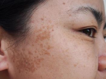
MELASMA
Finally, the most prominent risk factor for melasma onset and exacerbation is sun exposure. One small cohort study looking at Puerto Rican women found that in all cases, their mandibular melasma was exacerbated by sun exposure and lead to hyperpigmentation on histology.30 This finding suggests that populations closer to the equator, or more tropical conditions tend to have more risk factors for worsening melasma. Mahmoud and colleagues tested this concept and found that both visible light (400-700nm) and long- wavelength UVA1 (340-400nm) caused more prominent pigmentation in volunteers with darker skin (Fitzpatrick IV-VI), than those with skin type II, suggesting that even broader-spectrum sunscreens should be recommended, including those with iron oxide as a tint to help block visible light.31 Sun protection, thus, seems every bit as important for patients with skin of color: in addition to increasing the risk of skin cancer, sun exposure also plays a critical a role in pigmentary disorders.
VITILIGO
Whereas melasma represents the overproduction of pigmentation, vitiligo presents as the opposite problem: no pigmentation at all. Vitiligo affects about 1% of the population, incidence does not vary by skin color, and it affects men and women equally.32 The pathogenesis of vitiligo remains controversial, however a common theory supports an autoimmune origin since many patients with vitiligo tend to be diagnosed with autoimmune diseases, especially those involving the thyroid33. These factors add to the burden of disease, which has been studied in patients with vitiligo. A national study by American Academy of Dermatology was completed using 2013 claims data to examine the burden of disease in various skin diseases. The medical costs for vitiligo were calculated at $49 million and lost productivity of $6 million.34 Although this was on the lower end of the spectrum compared to the other skin diseases studied, it still demonstrates the financial burden posed by this disease. Patients with darker skin types tend to fail conservative treatments and are more likely to need phototherapy, which can be more expensive and time consuming.35
That being said, some may argue that the psychosocial burden of vitiligo is even more salient. Across ethnicities, patient vignettes on the effect of this disease show themes of low self-esteem, grief, and humiliation that affect every day life. There is not a consensus on the effect of skin color on psychosocial burden from vitiligo. Several studies demonstrate that patients with darker skin types (Middle Eastern, Caribbean and Indian heritage) perceived greater burden compared to lighter-skinned individuals 36, and another claimed that African Americans experience loss of racial identity37, yet the overall stress associated with vitiligo was the same across ethnic groups in more recent works.38 The burden of disease in vitiligo is significant across every group, and dermatologists need to keep psychosocial factors in mind when treating all patients with vitiligo.
UNITED COLORS OF DERMATOLOGY
There are important differences affecting people of color that need to be addressed and explored. Ideally, dermatologists will unite around their patients by continuing public education on skin exams and skin protection in various languages, and working with primary care colleagues to enhance screening and referrals. Increasing diversity among practicing dermatologists may also help to combat these disparities. While patients may feel more comfortable and increasing diversity in medicine will likely help, until that time, it is important to be aware of these disparities and important differences.
Any dermatologist can become more knowledgeable about these issues by joining the
References
1. Kaidbey KH, Agin PP, Sayre RM, Kligman AM. Photoprotection by melanin-a comparison of black and Caucasian skin. Journal of the American Academy of Dermatology. 1979;1(3):249-260. doi:10.1016/S0190-9622(79)70018-1
2. Chiesa Fuxench ZC, Block J, Boguniewicz M, et al. Atopic Dermatitis in America Study: a cross-sectional study examining the prevalence and disease burden of atopic dermatitis in the US adult population. Journal of Investigative Dermatology. October 2018. doi:10.1016/j.jid.2018.08.028
3. Hanifin JM, Reed ML. A population-based survey of eczema prevalence in the United States. Dermatitis: Contact, Atopic, Occupational, Drug. 2007;18(2):82-91.
4. Kaufman BP, Guttman-Yassky E, Alexis AF. Atopic dermatitis in diverse racial and ethnic groups-Variations in epidemiology, genetics, clinical presentation and treatment. Experimental Dermatology. 2018;27(4):340-357. doi:10.1111/exd.13514
5. Janumpally SR, Feldman SR, Gupta AK, Fleischer AB. In the United States, Blacks and Asian/Pacific Islanders Are More Likely Than Whites to Seek Medical Care for Atopic Dermatitis. Arch Dermatol. 2002;138(5):634-637. doi:10.1001/archderm.138.5.634
6. Sanyal RD, Pavel AB, Glickman J, et al. Atopic dermatitis in African American patients is TH2/TH22-skewed with TH1/TH17 attenuation. Annals of Allergy, Asthma & Immunology. September 2018. doi:10.1016/j.anai.2018.08.024
7. Noda S, Suárez-Fariñas M, Ungar B, et al. The Asian atopic dermatitis phenotype combines features of atopic dermatitis and psoriasis with increased TH17 polarization. J Allergy Clin Immunol. 2015;136(5):1254-1264. doi:10.1016/j.jaci.2015.08.015
8. Fischer AH, Shin DB, Margolis DJ, Takeshita J. Racial and ethnic differences in health care utilization for childhood eczema: An analysis of the 2001-2013 Medical Expenditure Panel Surveys. J Am Acad Dermatol. 2017;77(6):1060-1067. doi:10.1016/j.jaad.2017.08.035
9. SEER Cancer Statistics Review 1975-2004 - Previous Version - SEER Cancer Statistics. SEER. https://seer.cancer.gov/archive/csr/1975_2004/index.html. Accessed November 4, 2018.
10. Rouhani P, Hu S, Kirsner RS. Melanoma in Hispanic and Black Americans. Cancer Control. 2008;15(3):248-253. doi:10.1177/107327480801500308
11. Dawes SM, Tsai S, Gittleman H, Barnholtz-Sloan JS, Bordeaux JS. Racial disparities in melanoma survival. Journal of the American Academy of Dermatology. 2016;75(5):983-991. doi:10.1016/j.jaad.2016.06.006
12. Cormier JN, Xing Y, Ding M, et al. Ethnic differences among patients with cutaneous melanoma. Archives of Internal Medicine. 2006;166(17):1907-1914. doi:10.1001/archinte.166.17.1907
13. Wu X-C, Eide MJ, King J, et al. Racial and ethnic variations in incidence and survival of cutaneous melanoma in the United States, 1999-2006. Journal of the American Academy of Dermatology. 2011;65(5, Supplement 1):S26.e1-S26.e13. doi:10.1016/j.jaad.2011.05.034
14. Amber KT, Bloom R, Abyaneh M-AY, et al. Patient Factors and Their Association with Nonmelanoma Skin Cancer Morbidity and the Performance of Self-skin Exams. J Clin Aesthet Dermatol. 2016;9(9):16-22.
15. Coups EJ, Stapleton JL, Hudson SV, Medina-Forrester A, Goydos JS, Natale-Pereira A. Skin Cancer Screening Among Hispanic Adults in the United States: Results From the 2010 National Health Interview Survey. Arch Dermatol. 2012;148(7):861-863. doi:10.1001/archdermatol.2012.615
16. Van Voorhees AS, Enos CW. Diversity in Dermatology Residency Programs. Journal of Investigative Dermatology Symposium Proceedings. 2017;18(2):S46-S49. doi:10.1016/j.jisp.2017.07.001
17. Brown TJ, Rosen T, Orengo IF. Hidradenitis suppurativa. South Med J. 1998;91(12):1107-1114.
18. Lee DE, Clark AK, Shi VY. Hidradenitis Suppurativa: Disease Burden and Etiology in Skin of Color. DRM. 2017;233(6):456-461. doi:10.1159/000486741
19. Reeder VJ, Mahan MG, Hamzavi IH. Ethnicity and Hidradenitis Suppurativa. Journal of Investigative Dermatology. 2014;134(11):2842-2843. doi:10.1038/jid.2014.220
20. Vaidya T, Vangipuram R, Alikhan A. Examining the race-specific prevalence of hidradenitis suppurativa at a large academic center; results from a retrospective chart review. Dermatology Online Journal. 2017;23(6):3.
21. Deckers IE, Janse IC, van der Zee HH, et al. Hidradenitis suppurativa (HS) is associated with low socioeconomic status (SES): A cross-sectional reference study. Journal of the American Academy of Dermatology. 2016;75(4):755-759.e1. doi:10.1016/j.jaad.2016.04.067
22. Homma H. On Apocrine Sweatglands in White and Negro Men and Women. Bulletin of the Johns Hopkins Hospital. 1926;38(5). https://www.cabdirect.org/cabdirect/abstract/19272900338. Accessed November 8, 2018.
23. Thomas C, Rodby KA, Thomas J, Shay E, Antony AK. Recalcitrant Hidradenitis Suppurativa: An Investigation of Demographics, Surgical Management, Bacterial Isolates, Pharmacologic Intervention, and Patient-reported Health Outcomes. Am Surg. 2016;82(4):362-368.
24. Handog EB, Enriquez-Macarayo MJ. Melasma and Vitiligo in Brown Skin. Springer; 2017.
25. Handel AC, Miot LDB, Miot HA. Melasma: a clinical and epidemiological review. An Bras Dermatol. 2014;89(5):771-782. doi:10.1590/abd1806-4841.20143063
26. Pandya AG, Guevara IL. DISORDERS OF HYPERPIGMENTATION. Dermatologic Clinics. 2000;18(1):91-98. doi:10.1016/S0733-8635(05)70150-9
27. Wong RC, Ellis CN. Physiologic skin changes in pregnancy. Journal of the American Academy of Dermatology. 1984;10(6):929-940. doi:10.1016/S0190-9622(84)80305-9
28. Ortonne JP, Arellano I, Berneburg M, et al. A global survey of the role of ultraviolet radiation and hormonal influences in the development of melasma. Journal of the European Academy of Dermatology and Venereology. 2009;23(11):1254-1262. doi:10.1111/j.1468-3083.2009.03295.x
29. Pawaskar MD, Parikh P, Markowski T, McMichael AJ, Feldman SR, Balkrishnan R. Melasma and its impact on health-related quality of life in Hispanic women. The Journal Of Dermatological Treatment. 2007;18(1):5-9.
30. Pagán: Mandibular melasma - Google Scholar. https://scholar-google-com.proxy.uchicago.edu/scholar_lookup?title=Clinical%20studies%2C%20Mandibular%20melasma&author=RM.%20Pagan&author=JL.%20Sanchez&journal=PRHSJ&volume=19&issue=3&pages=231-4&publication_year=2000. Accessed November 8, 2018.
31. Mahmoud BH, Ruvolo E, Hexsel CL, et al. Impact of long-wavelength UVA and visible light on melanocompetent skin. J Invest Dermatol. 2010;130(8):2092-2097. doi:10.1038/jid.2010.95
32. Alikhan A, Felsten LM, Daly M, Petronic-Rosic V. Vitiligo: A comprehensive overview: Part I. Introduction, epidemiology, quality of life, diagnosis, differential diagnosis, associations, histopathology, etiology, and work-up. Journal of the American Academy of Dermatology. 2011;65(3):473-491. doi:10.1016/j.jaad.2010.11.061
33. Baldini E, Odorisio T, Sorrenti S, et al. Vitiligo and Autoimmune Thyroid Disorders. Front Endocrinol (Lausanne). 2017;8. doi:10.3389/fendo.2017.00290
34. Lim HW, Collins SAB, Resneck JS, et al. The burden of skin disease in the United States. Journal of the American Academy of Dermatology. 2017;76(5):958-972.e2. doi:10.1016/j.jaad.2016.12.043
35. Gawkrodger DJ, Ormerod AD, Shaw L, et al. Guideline for the diagnosis and management of vitiligo. British Journal of Dermatology. 2008;159(5):1051-1076. doi:10.1111/j.1365-2133.2008.08881.x
36. Living with vitiligo: results from a national survey indicate differences between skin phototypes - Ezzedine - 2015 - British Journal of Dermatology - Wiley Online Library. https://onlinelibrary-wiley-com.proxy.uchicago.edu/doi/full/10.1111/bjd.13839. Accessed December 3, 2018.
37. Porter JR, Beuf AH. Racial variation in reaction to physical stigma: a study of degree of disturbance by vitiligo among black and white patients. J Health Soc Behav. 1991;32(2):192-204.
38. Grimes PE, Miller MM. Vitiligo: Patient stories, self-esteem, and the psychological burden of disease. Int J Womens Dermatol. 2018;4(1):32-37. doi:10.1016/j.ijwd.2017.11.005
39. Ethnic Skin Care | Skin of Color Dermatology. Skin of Color Society. http://skinofcolorsociety.org/about-socs/. Accessed November 4, 2018.
Articles in this issue
over 7 years ago
Early-stage melanoma surveillanceover 7 years ago
Two big trends in cosmeceuticals for 2019over 7 years ago
Initiative uses dermatology to address total women’s healthover 7 years ago
Over the counter options for foot dermatosesover 7 years ago
Fast, effective tool to diagnose nail fungusover 7 years ago
Fat intake linked to skin cancer riskover 7 years ago
How to manage practice changeover 7 years ago
Expect exciting trends in dermatology careover 7 years ago
New fungal medication for the U.S. marketAdvertisement
Related Content
Advertisement


Latest CME


Advertisement
Advertisement
Trending on Dermatology Times
1
Real-World Study Finds Experienced Dermatologists Outperform AI in Skin Cancer Diagnosis
2
New Review Confirms How Nonmedicated Skin Care Vehicles Drive Clinical Improvement
3
Zasocitinib Shows High Skin Clearance at Hard-to-Treat Psoriasis Sites
4
Dermatology Times Spot Test: July 19, 2026
5