
|Articles|February 12, 2020
- Dermatology Times, February 2020 (Vol. 41, No. 2)
- Volume 41
- Issue 2
How to diagnose rosacea in skin of color patients
Author(s)John Jesitus
Susan C. Taylor, M.D., shares distinguishing characteristics of differential diagnoses to keep in mind when examining skin of color patients for rosacea.
Advertisement
A paucity of studies and case reports has contributed to the misconception that rosacea rarely afflicts patients with skin of color (SOC), say authors of a recent review.
“Rosacea in SOC patients is grossly under-recognized and underdiagnosed,” says senior author Susan C. Taylor, M.D. “This is because it is often difficult for dermatologists and other providers to appreciate the erythema that is diagnostic.”1 She is associate professor of dermatology at the University of Pennsylvania’s Perelman School of Medicine, vice president-elect of the American Academy of Dermatology and founder of the Skin of Color Society.
In a separate publication, investigators using the search term “rosacea” found 3,786 articles indexed for MEDLINE, versus just 32 for the terms “rosacea” and “skin of color.”2
The true prevalence of rosacea in SOC remains unknown. While a 2018 meta-analysis estimated global rosacea prevalence in all adults to be 5.46%, estimates of rosacea prevalence in SOC patients in individual studies range from 0.4% (Angola) to 12.4% (Colombia).
Underdiagnosis and misdiagnosis of rosacea in SOC lead to increased morbidity.
“Symptoms of burning, stinging, erythema, papules and pustules negatively impact patient quality of life, and patients may suffer needlessly,” says Dr. Taylor.
Undiagnosed rosacea moreover may progress to disfiguring rhinophyma. Likewise, the fact that the granulomatous variant of rosacea has been predominantly reported in black patients may stem from increased susceptibility in these patients.3
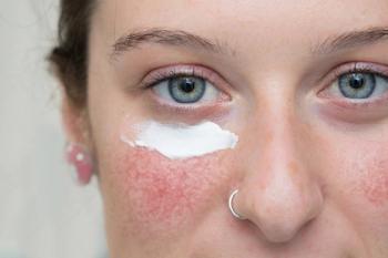
To highlight erythema and telangectasias in SOC, Taylor et al. recommend using adequate lighting, skin blanching and dermatoscopy.
“When you apply pressure to the skin with a slide or dermatoscope,” explains Dr. Taylor, “the blood will drain from the skin, and the skinappears white beneath the slide or dermatoscope. Then when you release the pressure, the blood comes back into the skin, and you can then appreciate the redness.”
Similarly, photographing the affected area against a dark blue background provides a contrast that makes identifying redness easier, she adds.
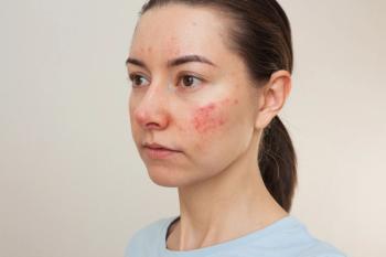
In the differential diagnosis of rosacea, systemic lupus erythematosus (SLE) occurs particularly more commonly in SOC, Dr. Taylor says. The malar or butterfly rash of SLE can appear similar to rosacea, but the malar rash typically spares the nasolabial folds, while rosacea does not. Conversely, papules and pustules rarely occur in SLE.
To confirm or rule out SLE, Taylor et al. suggest performing a complete physical examination and a skin biopsy with hematoxylin and eosin and direct immunofluorescence, as well as checking serologies including antinuclear antibody (ANA).
Although elevated ANA has been reported in patients with rosacea, it is lower than what is typically seen in SLE. If ANA is elevated, Dr. Taylor advises checking more specific SLE antibodies; for example, the double-stranded DNA test is positive in lupus. SLE also can be differentiated histologically by a considerably lower CD4:CD8 ratio, fewer CD4+CD25+ regulatory T cells and more CD123+ plasmocytoid dendritic cells versus rosacea.
Rosacea and seborrheic dermatitis can present concurrently. Symptoms of seborrheic dermatitis include erythematous patches and plaques involving the scalp, anterior and posterior hairlines, pre- and post-auricular areas and medial eyebrows. Both rosacea and seborrheic dermatitis may impact the nasolabial folds, but the presence of scale distinguishes the latter.
Additionally, the erythematous lesions of seborrheic dermatitis are often annular, and the postinflammatory hypopigmentation (and to a lesser extent, hyperpigmentation) common to this condition are relatively uncommon in rosacea.
The heliotrope rash that marks dermatomyositis differs from rosacea in its dusky, violaceous hue and its periorbital involvement. Other signs that indicate dermatomyositis, which is four times more common in African-American versus Caucasian patients, include edema of the face and extremities, Gottron papules and poikiloderma.
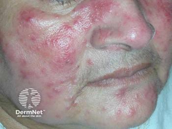
Cutaneous sarcoidosis may present with multiple morphologic features, most typically firm yellow, brown, violaceous, red or flesh-colored monomorphic papules or nodules affecting the perioral, periocular, medial and/or lateral face. However, plaques, lupus pernio, subcutaneous infiltrates and infiltrates of scars also have been reported. Sarcoid papules typically measure 1 to 5 mm on the face, neck and periorbital skin. They are initially orange or yellow-brown, then turn brownish-red or violaceous before involuting to form faint macules. Papular lesions also may evolve into plaques, particularly on the extremities, face, scalp, back and buttocks.
Unlike sarcoidosis, granulomatous rosacea lacks plaques, lupus pernio, subcutaneous infiltrates and infiltration of scars. Although patients with granulomatous rosacea may report pain, pruritus or burning, these patients do not experience the ushing and erythema seen in more typical rosacea presentations.
Distinguishing features of steroid dermatitis may include full-facial involvement, versus rosacea’s centrofacial predilection. Regarding acne vulgaris, patients with this condition do not experience telangectasias and flushing, while those with rosacea do not experience comedonal lesions but can have hyperpigmented macules.
Disclosures:
Dr. Taylor has been an investigator, speaker and advisory board member for Aclaris (maker ofoxymetazoline) and an advisory board member for Galderma (maker of brimonidine).
References:
1. Onalaja AA, Lester JC, Taylor SC. Establishing the diagnosis of rosacea in skin of color patients. Cutis. 2019;104:38-41.
2. Alexis AF. Rosacea in patients with skin of color: uncommon but not rare. Cutis. 2010;86:60-62.
3. Dlova NC, Mosam A. Rosacea in black South Africans with skin phototypes V and VI. Clin Exp Dermatol. 2017;42:670-673.
Articles in this issue
over 6 years ago
Gene expression profile testing aids skin cancer managementover 6 years ago
Rosacea commonly underdiagnosedover 6 years ago
Technology targets non-genital wartsover 6 years ago
Can sweat shed a light on disease severity in atopic dermatitis?over 6 years ago
Adjunctive platelet-rich plasma may improve acne scarsover 6 years ago
An investigational drug for keloidsover 6 years ago
Study supports evidence on indoor tanning riskover 6 years ago
Nano-pulse stimulation promising for skin lesionsover 6 years ago
Mind-body therapies effective for skin disordersover 6 years ago
Dermatologist closes gaps for patients with skin of colorAdvertisement
Related Content
Advertisement


Latest CME


Advertisement
Advertisement
Trending on Dermatology Times
1
Stapokibart Successfully Treats Refractory CSU After Omalizumab Failure in New Case Report
2
Single-Dose IL-2 Fusion Protein Shows Hair Regrowth Signal in Early Trial
3
Oral STAT6 Degrader KT-621 Delivers Biologics-Like Efficacy in Moderate to Severe AD
4
Study Finds Psoriasis Patients Trust Topical Therapies Less Than Those With AD
5