Red Fluorescence Highlights Microbial Composition Variability in Androgenetic Alopecia
Researchers noted differences in microbial evenness, abundance composition, and functional prediction.
Ultraviolet-induced red fluorescence dermoscopy (UVFD) is capable of highlighting red fluorescence and microbial composition variability in patients with androgenetic alopecia (AGA), according to findings from a
Researchers noted significant differences in microbial evenness, abundance composition, and functional predictions across different regions of the scalp. To their knowledge, this is the first known study to investigate scalp red fluorescence in the microbiological mechanisms of AGA.
Background and Methods
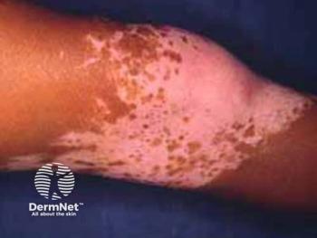
Ultraviolet (UV)-induced fluorescence is used in disease diagnosis of vitiligo, scabies, and pityrosporum folliculitis, for example. Similar to the diagnostic tool used in vitiligo, Wood's lamp examination illuminates pityrosporum folliculitis, highlighting blue-white fluorescence; this tool provides a visual differentiation between this form of folliculitis and another, bacterial folliculitis.2
Researchers Zhang et al noted that while fluorescence has been used to identify scalp conditions such as tinea capitis, its use has not been explored in AGA. A 2018 study published in Dermatology found that when exposed to UV light, microbes are capable of emitting specific fluorescence. Human follicular ultraviolet-induced red fluorescence (UVRF) is emitted due to the presence of native bacteria as opposed to sebum, they reported.3
In the present study, researchers conducted a study of 36 adult patients ages 18 to 60 years with AGA diagnosed between November 2022 and April 2023. Qualifying criteria included age and presence of fluorescent and non-fluorescent areas of AGA on the scalp. Exclusionary criteria included the presence of existing systemic or scalp-related conditions, the use of antibiotics, glucocorticoids, or live bacterial formulations within 3 months from the start of the study, and individuals who were lactating or pregnant were unable to participate.
Researchers collected clinical photographs and UV dermatoscope imaging of all patients and collected scalp samples from both affected and non-affected areas. Samples were placed in sterile tubes and stored for later DNA extraction.
Microbial DNA from scalp swab samples was extracted using the CTAB method, assessed by gel electrophoresis, and quantified with a UV spectrophotometer. The V3-V4 regions of the 16S rRNA gene were PCR-amplified and sequenced on the NovaSeq 6000 system. Bioinformatics analysis involved quality filtering, removing chimeras, and taxonomic classification using the SILVA database. Alpha and beta diversity were analyzed with QIIME2 and visualized in R. Functional predictions were made with PICRUSt2, and statistical analyses used STAMP. Swabs cultured on agar plates were identified using MALDI-TOF MS, focusing on red-fluorescent colonies with confidence scores ≥1.7.
Findings
UVFD examination revealed distinct regions of fluorescence and non-fluorescence on the scalp. Areas showing red fluorescence contrasted significantly with non-fluorescent regions.
In analyzing scalp microbial diversity, results showed high sequencing coverage, indicating a thorough representation of the samples. The number of microbial species did not significantly differ between fluorescent and non-fluorescent areas. However, the evenness of species distribution was lower in the fluorescent group.
The composition of bacterial communities was compared at both phylum and genus levels. Actinobacteriota, firmicutes, and proteobacteria were the most prevalent phyla, with significant differences in their abundance between In analyzing scalp microbial diversity, results showed high sequencing coverage, indicating a thorough representation of the samples.
The number of microbial species did not significantly differ between fluorescent and non-fluorescent areas. Actinobacteriota were more abundant in fluorescent regions, whereas proteobacteria were more common in non-fluorescent regions. At the genus level, cutibacterium and staphylococcus were predominant, with significant variations in their relative abundances between the 2 groups.
Cultivation of scalp swab bacteria under both anaerobic and aerobic conditions identified several red fluorescent colonies. Anaerobic cultures predominantly yielded cutibacterium spp. and staphylococcus epidermidis, both showing distinct red fluorescence. Aerobic cultures revealed micrococcus spp. The fluorescence intensity of S. epidermidis notably increased upon air exposure, highlighting a dynamic change in fluorescence under different environmental conditions.
Conclusions
The study highlights the potential of UV light-induced fluorescence and other fluorescence-based techniques in dermatological diagnostics and treatment, particularly concerning scalp health. The findings suggest that red fluorescence could indicate inflammation, which is prevalent around miniaturized hair follicles in AGA.
The study has limitations, such as the inconsistent literature on facial and hair follicle red fluorescence and its relationship with sebum secretion. The homogeneity in the participants' racial backgrounds and living environments also limits the generalizability of the findings.
According to study authors, future studies should explore the specific wavelengths of red fluorescence emitted by microbial metabolites and their implications for scalp health.
"Scalp microbiota are intimately associated with scalp health," wrote Zhang et al. "The observed red fluorescence should not be overlooked, as continued exploration of this phenomenon will undoubtedly enhance our understanding of scalp health."
References
- Zhang L, Hu Y, Xie B, et al. Ultraviolet-induced red fluorescence in androgenetic alopecia—indicating alterations in microbial composition. Skin Res Technol. Published online June 20, 2024.
https://doi.org/10.1111/srt.13777 - Klatte JL, van der Beek N, Kemperman PM. 100 years of Wood's lamp revised. J Eur Acad Dermatol Venereol. 2015; 29(5): 842-847. doi:
10.1111/jdv.12860 - Xu DT, Yan JN, Liu W, et al. Is human sebum the source of skin follicular ultraviolet-induced red fluorescence? A cellular to histological study. Dermatology. 2018; 234(1-2): 43-50. doi:
10.1159/000489396
Newsletter
Like what you’re reading? Subscribe to Dermatology Times for weekly updates on therapies, innovations, and real-world practice tips.


