Method may allow 'optical' biopsy
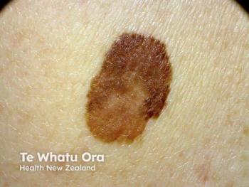
New technology developed by a University of Rochester (N.Y.) optics professor may enable doctors to examine potentially cancerous skin lesions without having to physically biopsy them, ScienceDaily.com reports.
Rochester, N.Y. - New technology developed by a University of Rochester (N.Y.) optics professor may enable doctors to examine potentially cancerous skin lesions without having to physically biopsy them, ScienceDaily.com reports.
Jannick Rolland, Ph.D., professor of biomedical engineering at the university’s Institute of Optics, led a team of researchers in developing a unique liquid-lens setup for a process called optical coherence microscopy.
Using this technology, the tip of a cylindrical probe is placed in contact with the suspect tissue. As the electrical field around the water-droplet lens changes, the droplet changes shape, thereby changing the focus of the lens.
This allows the taking of thousands of pictures at different depths below the skin’s surface. Combining these images creates a precisely focused image of all the tissue structure up to 1 mm deep.
ScienceDaily.com quotes Dr. Rolland as saying, “My hope is that in the future, this technology could remove significant inconvenience and expense from the process of skin lesion diagnosis. When a patient walks into a clinic with a suspicious mole, for instance, they wouldn’t have to have it necessarily surgically cut out of their skin or be forced to have a costly and time-consuming MRI done.
“Instead, a relatively small, portable device could take an image that will assist in the classification of the lesion right in the doctor’s office.”
Because the device uses near-infrared light instead of ultrasound, image resolution is micron-scale as opposed to millimeter-scale - thus allowing for a high-resolution, 3-D image of what lies below the skin’s surface.
The process has been tested successfully in vivo, and several papers on it have been published in peer-reviewed journals, according to ScienceDaily.com. Plans call for the device to be used in clinical research to assess its ability to discriminate between different types of lesions.
Dr. Rolland presented her findings at the annual meeting of the American Association for the Advancement of Science, held last month in Washington.
Newsletter
Like what you’re reading? Subscribe to Dermatology Times for weekly updates on therapies, innovations, and real-world practice tips.


