|Articles|September 1, 2010
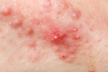
Giant congenital nevi may pose threat of melanoma
If congenital nevi are small, there is insufficient evidence to suggest they should be removed, for they are not likely to transform into melanoma, according to the chief dermatologist at Phoenix Children's Hospital, Phoenix. However, giant nevi present a much higher risk, says Ron Hansen, M.D., F.A.A.D., a board-certified dermatologist and board-certified pediatrician.
Advertisement
Key Points
Phoenix - If congenital nevi are small, there is insufficient evidence to suggest they should be removed, for they are not likely to transform into melanoma, according to the chief dermatologist at Phoenix Children's Hospital, Phoenix.
"Congenital nevi are quite common," Dr. Hansen says. "Up to 2 percent of children can have a small congenital nevus. There is a very low risk of children developing melanoma if they have a small congenital nevus."
Small congenital nevi measure less than 2 cm, and if they do transform into melanoma, it is usually detectable by clinicians, and the melanoma develops at puberty or later, Dr. Hansen says.
"At one point, I was more enthusiastic about excising (small congenital nevi), but statistically, you would have to treat about 1,000 cases to avoid one case of melanoma," he says.
"There is usually a spot that presents within the nevus," Dr. Hansen says. "In those cases, you would see the melanoma coming, so it would be salvageable. You can identify the melanoma by a color change. It's not common sense to remove the small congenital nevi."
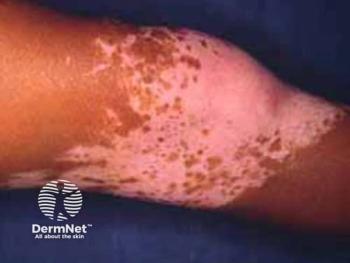
By contrast, when infants present with giant congenital nevi, the lesions pose a much higher risk, according to Dr. Hansen.
"Such lesions would carry a risk as high as 5 percent, in terms of the development of a melanoma," says Dr. Hansen, noting a common site where giant nevi present would be on the back.
"When a giant nevus presents in newborns, it would measure more than 6 cm, and in adolescents, they would measure more than 20 cm."
Looking deeper
"The risk of melanoma is directly related to how deep the melanoma measures, and, in giant nevi, they often arise deep in the nevus," Dr. Hansen says.
Hence, melanomas that develop in giant congenital nevi are deep, difficult to excise, and likely to be metastatic, Dr. Hansen explains. "They are worrisome, and are not readily recognized by surface changes," he says.
Another factor that influences the risk of complication with a giant nevus is the presence of satellite nevi, which usually accompany a giant nevus, but are substantially smaller in size.
"If a patient has a huge nevus and also many satellite lesions, and the nevus reaches the scalp from the back, there is a risk of leptomeningeal melanosis," Dr. Hansen warns. He notes that children who develop leptomeningeal melanosis may face "devastating" neurological complications.
Newsletter
Like what you’re reading? Subscribe to Dermatology Times for weekly updates on therapies, innovations, and real-world practice tips.
Advertisement
Related Content
Advertisement


Latest CME


Advertisement
Advertisement
Trending on Dermatology Times
1
FDA Accepts Galderma’s BLA Resubmission for Liquid Neuromodulator RelabotulinumtoxinA
2
Icotrokinra Shows Superior Efficacy Over Advanced Oral Therapies in New Psoriasis Meta-Analysis
3
MAIC Evaluates 1-Year Outcomes of Biologics in HS
4
Introducing Dermatology Times NP/PA Connect
5