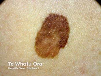
FISH helps evaluate Spitzoid tumors
Fluorescence in situ hybridization (FISH) analysis may improve the prognostic evaluation of atypical Spitzoid tumors, results of a new study indicate, HealthDay News reports.
Florence, Italy - Fluorescence in situ hybridization (FISH) analysis may improve the prognostic evaluation of atypical Spitzoid tumors, results of a new study indicate, HealthDay News reports.
Working with a group of 38 patients, researchers from the University of Florence evaluated whether the presence of chromosomal abnormalities could assist with diagnostic and prognostic assessment of atypical Spitzoid lesions with a thickness of 1 mm or more. When the patients’ clinicopathological features were analyzed, chromosomal abnormalities were identified by FISH analysis in 25 patients. The mutational status of BRAFV600E and H-RAS genes was assessed by allele-specific real-time polymerase chain reaction and direct sequencing, respectively.
The researchers identified micrometastases in four of the nine patients who had a sentinel lymph node biopsy. In those who did not undergo biopsy, four had bulky lymph node metastases and one experienced visceral metastases and death. Patients with lymph node involvement showed significantly more deep mitoses, less inflammation and more plasma cells in Spitzoid lesions. Chromosomal alterations were detected by FISH analysis in six patients. Follow-up data from the FISH analysis indicated that the only fatal outcome exhibited multiple chromosomal alterations. BRAFV600E mutation was detected in 75 percent of cases and H-RAS mutation on exon 3 was detected in 27 percent of cases.
“After a careful histologic evaluation, FISH analysis may be of help for diagnosis and prognosis,” the authors wrote, adding that results are preliminary but justify further research and follow-up.
The study, which appears in the May issue of the Journal of the American Academy of Dermatology, was partially funded by Abbott Molecular.
Newsletter
Like what you’re reading? Subscribe to Dermatology Times for weekly updates on therapies, innovations, and real-world practice tips.


