

Quiz|Articles|May 9, 2026
In the Chair: Managing Nail Dystrophy in AD
Key Takeaways
- Atopic nail dystrophy can mimic onychomycosis, psoriasis, or lichen planus, so negative mycology and dermoscopic eczematous patterns are pivotal for accurate attribution to periungual AD.
- Periungual nail unit changes reflect Th2/JAK1-STAT3 inflammatory signaling, making antifungals ineffective when infection is excluded and reinforcing the nail as a downstream target of systemic control.
In the Chair puts you in the hot spot, challenging you to navigate complex, real-world cases.
Advertisement
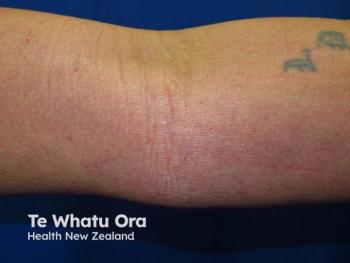
Nail dystrophy in atopic dermatitis is easy to miss — and easy to misattribute. When a patient with hand and foot eczema develops onycholysis and nail plate thickening, the first instinct is often to rule out onychomycosis and move on.1 But in patients with an atopic background, nail changes can be a direct extension of periungual inflammation, driven by the same Th2/JAK1 signaling that governs the rest of the disease. And they don’t behave like skin.
This case — published in Clinical, Cosmetic and Investigational Dermatology in 2026 — follows a 30-year-old woman with refractory acral eczema and progressive nail dystrophy across a 12-month treatment course with the selective JAK1 inhibitor abrocitinib.2 It raises 3 clinical decision points that APPs are increasingly likely to encounter as JAK inhibitor use in AD expands: how to establish the diagnosis, how to respond when initial dosing fails, and how to counsel patients when the skin clears before the nails do.
THE PATIENT
A 30-year-old woman presents with a 1-year history of recurrent eczematous lesions affecting both hands and feet, accompanied by progressive nail changes. The course is chronic and relapsing, predominantly acral, with frequent refractory flares. She reports a history of allergic rhinitis.
Previous diagnoses have included eczema, tinea pedis, and onychomycosis; she has been treated with oral antihistamines, itraconazole, multiple topical corticosteroids, and antifungal agents with limited and unsustained improvement.
On examination:
- Extensive eczematous dermatitis of the hands and feet
- Erythema, vesiculation, fissuring, and marked pruritus
- Prominent nail involvement:
- Onycholysis
- Nail plate thickening
- Surface roughness
- Involvement affecting:
- Left great toenail
- Left middle fingernail
- Left little fingernail
Baseline Disease Activity:
- SCORAD: 47
- PP-NRS: 9
- Sleep disturbance score: 9
CLINICAL DECISION POINT 1 — Establishing the Diagnosis
Before initiating systemic therapy, you complete a targeted workup.
Results
- Routine biochemical tests: Normal
- Thyroid function tests: Normal
- Autoimmune parameters: Normal
- Serum total IgE: Mildly elevated at 135.4 kIU/L
- Fungal examination: Negative
- Dermoscopy: Consistent with an eczematous process
⚠ Diagnostic Consideration
Onychomycosis, psoriatic nail disease, and lichen planus are the most clinically relevant mimics of eczematous nail dystrophy.
Negative fungal exam, absence of the “oil drop” sign or geometric pitting associated with psoriasis, and dermoscopic findings consistent with an eczematous process — rather than fungal or psoriatic changes — support an AD-associated etiology in this patient.
📋 WHAT WOULD YOU DO?
Fungal exam is negative. IgE is mildly elevated. Dermoscopy suggests an eczematous process. The patient has a history of allergic rhinitis and refractory acral eczema. How do you establish the primary diagnosis driving the nail changes?
CLINICAL DECISION POINT 2 — When the First Dose Doesn’t Work
Given the chronic refractory course, inadequate response to conventional treatments, and known difficulty of treating acral and periungual disease with topicals, systemic targeted therapy is initiated.
Initial Treatment:
- Abrocitinib 100 mg once daily
At 4 Weeks:
- No meaningful clinical improvement
- Severe pruritus persists
- Nail thickening progresses
📋 WHAT WOULD YOU DO?
Your patient has shown no meaningful response to abrocitinib 100 mg at 4 weeks. Pruritus remains severe; nail thickening has progressed. What is your next step?
CLINICAL DECISION POINT 3 — The Skin Has Cleared, But the Nails Haven’t
By month 4, hand and foot eczema has almost completely resolved. However, nail morphology, though improved, continues to show onycholysis and surface irregularity.
The patient asks whether the nails will ever fully recover and whether the abrocitinib is still working.
⚠ Clinical Clue:
- Fingernails grow approximately 3 mm per month
- Toenails grow approximately 1.5 mm per month
- Full nail plate replacement takes:
- ~6 months for fingernails
- Up to 12–18 months for toenails
Nail structural recovery after inflammatory nail disease reflects regrowth from a healing matrix — not delayed anti-inflammatory activity.
📋 WHAT WOULD YOU DO?
At month 4, eczema has cleared but nail morphology remains abnormal. The patient is concerned. How do you counsel her and what, if anything, do you adjust?
12-MONTH TREATMENT TIMELINE
Timepoint
Dose
Cutaneous/Systemic Response
Nail Status
Baseline
Abrocitinib 100 mg
SCORAD 47; PP-NRS 9; sleep score 9
Severe pruritus persists; nail thickening progresses
Week 4
Abrocitinib 100 mg
No meaningful improvement
Dose escalated to 200 mg
Week 6 (+2 weeks)
Abrocitinib 200 mg
Marked pruritus relief; heel/plantar eczema improving
Minimal early toenail changes only
Month 4
Abrocitinib 200 mg
Hand/foot eczema near-complete clearance
Reduced onycholysis; smoother nail surface
Month 8
Abrocitinib 100 mg (tapered)
Cutaneous remission maintained
Continued nail structural recovery
Month 12
Abrocitinib 100 mg
Complete cutaneous remission; SCORAD near 0
Marked nail recovery; mild residual toenail thickening only; no adverse events
CLINICAL TAKEAWAYS
✓ AD-associated nail dystrophy is driven by periungual JAK1/STAT3-mediated inflammation — not infection
✓ Negative fungal exam and atopic background support the diagnosis
✓ Conventional topicals rarely reach the nail matrix
✓ Systemic targeted therapy is appropriate for refractory acral AD with nail involvement
✓ Dupilumab has been reported to paradoxically induce new-onset nail dystrophy in some AD patients
✓ JAK inhibitors may offer a more consistent anti-inflammatory effect on the nail unit
✓ Abrocitinib 100 mg may be escalated to 200 mg when adequate response is not achieved at 4 weeks
✓ Skin clearance precedes nail recovery by weeks to months — this reflects nail growth kinetics, not treatment failure
✓ Serial clinical photography is the most practical tool for tracking nail response
THE TAKEAWAY
Nail dystrophy in AD is underrecognized, poorly standardized, and therapeutically underdefined. This patient had been treated for onychomycosis she didn’t have, with antifungals that couldn’t address the periungual inflammation driving her nail changes.
The correct diagnosis was available once the fungal workup came back negative and the atopic background was taken seriously — but it required connecting the nail involvement to the eczema, not treating it as a separate problem.
The abrocitinib course in this case illustrates 2 things APPs need to manage proactively:
- Dose escalation at 4 weeks
- Lack of response to 100 mg is not necessarily treatment failure
- It may be a signal to escalate before switching therapeutic class
- Counseling during delayed nail recovery
- Skin may clear long before the nails normalize
- Patients need to understand the biology of nail regrowth
- Setting expectations improves adherence
As JAK inhibitor use in AD broadens, APPs will see more of this phenotype. Recognizing the nail unit as a downstream target of JAK1/STAT3 dysregulation — and setting appropriate expectations for recovery — is increasingly valuable clinical knowledge.
References
- Chung BY, Choi YW, Kim HO, Park CW. Nail dystrophy in patients with atopic dermatitis and its association with disease severity. Ann Dermatol. 2019;31(2):121-126. doi:10.5021/ad.2019.31.2.121
- Ma A, Deng Y. Effectiveness of abrocitinib in atopic dermatitis presenting with hand-foot eczema and nail dystrophy: a case report. Clin Cosmet Investig Dermatol. 2026;19:596189. Published 2026 May 1. doi:10.2147/CCID.S596189
Advertisement
Related Content
Advertisement


Latest CME


Advertisement
Advertisement
Trending on Dermatology Times
1
Visible Vitiligo Linked to Significant Stigma and Behavioral Adaptations, According to Global Survey
2
The New Economics of Dermatology Claims: Understanding and Responding to Rising Denials
3
Striking the Balance with JAK Inhibitors in Atopic Dermatitis Management
4
Ycanth Phase 3 Trial Dosing Begins for Common Warts in US, Japan
5