
|Articles|January 29, 2014
Digital skin lesion analysis may help dermatologists classify skin lesions
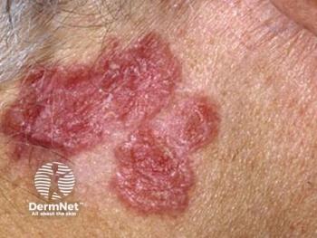
A multispectral digital skin lesion analysis device may be useful in helping clinicians to classify clinically ambiguous lesions, detect early melanomas in young adults, and identify outlier lesions that should be biopsied, according to a collection of posters presented at MauiDerm 2014.
Advertisement
A multispectral digital skin lesion analysis device (MSDSLA, MelaFind; MELA Sciences) may be useful in helping clinicians to classify clinically ambiguous lesions, detect early
The first study investigated the use of the device in young adults. Dr. Kollmann reviewed data for 183 lesions in 136 young adults ages 16 to 25 who had been enrolled in a pivotal study, which has been
Also included was real-world data from two clinical cases: a 17-year-old female and a 24-year-old male, as well as six invasive melanomas in young adults who had participated in the pivotal trial and were randomly selected to be reviewed by 211 American and German dermatologists as part of a reader study.
The 17-year-old patient was clinically unsuspicious for more than three years. Use of the MSDSLA reported a 3.5 score and pathology results indicated melanoma in situ. The 24-year-old patient had a lesion that appeared within four months of excision and grew. He was clinically diagnosed with a Spitz/Reed Nevus. Use of the MSDSLA reported a score of 3.3 and pathology results indicated melanoma in situ.
Next: Analysis results
More on melanoma detection
Of the 183 lesions examined in the pivotal study, 13 were high-grade melanoma lesions. MSDSLA correctly considered 11 of these lesions as possessing "high disorganization."
Average false negative rates for the 211 American and German dermatologists for the six melanomas reviewed in the reader study were: 48.6 percent (0.17 mm median Breslow thickness), 57.3 percent (0.60 mm median Breslow thickness), 1.9 percent (0.32 mm median Breslow thickness), 76.2 percent (0.15 mm median Breslow thickness), 46.0 percent (0.70 mm median Breslow thickness), and 78.1 percent (0.28 mm median Breslow thickness).
"MSDSLA can be used to help dermatologists decide which lesions may be suspicious for melanoma by providing information about the structure of a lesion from under the skin," Dr. Kollmann reported in the abstract.
In a second abstract, researchers looked retrospectively at MSDSLA-computed classifier scores pigmented lesions relative to clinical characteristics and histopathology. The researchers noted a range of scores between -5.2 to 9.0. Average classifier scores for melanomas, high grade lesions, nonmelanoma skin cancers, and benign lesions were 3.5, 2.7, 2.6 and 1.7 respectively, further validating use of the device.
A third abstract investigated whether the MSDSLA scores can be useful in identifying benign nevi that should be further evaluated. Noting that a pivotal study indicated that, in patients with multiple nevi, scores for melanoma and high grade lesions tend to be higher than benign lesions, researchers included 10 patients with at least 200 nevi and analyzed MSDSLA score distributions for each patient and for all patients.
The researchers found significantly different score distributions among some patients. Individual thresholds ranged from 2.8 to 4.5 with 76 percent of lesions falling above the zero threshold used for the general population and 2.1 percent falling above the individual thresholds calculated for each patient. The authors concluded that the analysis of MSDSLA scores can be used to identify typical lesions on a patient and outliers, which should then be considered for biopsy.
More on melanoma detection
Advertisement
Related Content
Advertisement


Latest CME


Advertisement
Advertisement
Trending on Dermatology Times
1
What Dermatologists Need to Know About the First New Sunscreen Ingredient in Decades
2
The Expanding Role of Immunotherapies for Non-Melanoma Skin Cancers
3
KT-621 in Atopic Dermatitis: What Early EASI and Pruritus Data Show
4
Filling the HS Treatment Gap: Ruxolitinib Targets Early-Stage Disease
5