

|Articles|October 8, 2019
- Dermatology Times, October 2019 (Vol. 40, No. 10)
- Volume 40
- Issue 10
Radiodermatitis increasingly common in female breast cancer patients
Author(s)Ilya Petrou, M.D.
Dermatologists investigate better ways to treat radiodermatitis as it becomes increasingly common in breast cancer patients.
Advertisement
With more female breast cancer survivors living longer,
In a commentary published in Breast Cancer - Targets and Therapy, a team led by Rene-Jean Bensadoun, M.D., a radiation oncologist with the Centre de Haute Energie in France, discussed not only how providers can recognize the types of radiodermatitis, but also some treatment options.
“Radiodermatitis is still a pregnant problem for patients receiving radiation for cancer,” he says. “Radiation oncology is having brilliant results, but acute and late skin toxicity remains an inherent side effect of most radiation treatments, especially in breast cancer, gynecological and rectal cancers.”
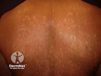
In female breast cancer patients, radiodermatitis presents in acute and chronic ways. Not only can these forms present differently, but therapeutic options also vary, he says.
ACUTE DERMATITIS
Acute dermatitis is identified by symptoms that appear within six months of treatment. Patients experience dry, itchy skin that can be red, feel warm, and peel along the skin folds of the breasts, clavicle, and periauricular areas.
To date, there have been few evidence-based recommendations on preventing acute radiodermatitis. However, Bensadoun says, new data points to dermocosmetics.
“The role of dermocosmetics has to be highlighted in the preventive management of these skin toxicities,” he says. “Previously, they were not prescribed by most physicians, but recent international guidelines have highlighted their important role.”
For treatment, he says, lasers are preferred. Both animal and clinical studies suggest low level light therapy (LLLT), also known as photobiomodulation or “soft laser” (red light or infrared with <150 mW power), can reduce inflammation and prevent fibrosis.
Bensadoun’s group recommends initiating this treatment after a few radiation sessions and before significant redness appears, if possible. However, don’t forego treatment if redness already exists.
CHRONIC DERMATITIS
With similar symptoms, chronic dermatitis appears after six months post-radiation treatment and up to 30 years later. The average is after 10-to-15 years. Subsequent chemotherapy or antibiotic treatment, repetitive trauma, sun exposure, or further irradiation are common prompts.
Research reveals pulsed dye laser treatment can clear telangiectasia, red streaks caused by dilated blood vessels. Using short 0.45-millisecond pulses with a 585-nm pulsed dye laser can whiten lesions. Data indicates three sessions can produce 90-percent lesion-size reduction with blood spots disappearing within 10-to-15 days. By comparison, intense pulsed light results in a 50% lesion reduction.
Ultimately, Bensadoun says, it’s critical for radiation oncologists and dermatologists to understand the frequency of radiodermatitis and how to ameliorate it. Â
References:
1. Seité S, Bensadoun RJ, Mazer JM. Prevention and treatment of acute and chronic radiodermatitis. Breast Cancer (Dove Med Press). 2017;9:551-557.
Articles in this issue
over 6 years ago
Non-melanoma skin cancer increases in the elderlyover 6 years ago
Looming medical billing and coding changes in healthcare policyalmost 7 years ago
7 tips to get the most from your social media postsalmost 7 years ago
IL-17 inhibitors may fill treatment gap in rosaceaalmost 7 years ago
Head and neck melanoma characteristicsalmost 7 years ago
Do continuing medical education certifications improve patient safety?Advertisement
Related Content
Advertisement


Latest CME


Advertisement
Advertisement
Trending on Dermatology Times
1
GX-03 Phase 2 Program Expands to Full Atopic Dermatitis Spectrum
2
Teva Advances TEV-'408 Into Phase 2b for Vitiligo
3
Topical GX-03 Shows Early Promise in Mild to Moderate Atopic Dermatitis
4
First-in-Class Oral Piclidenoson Reaches Pivotal Phase 3 Enrollment Milestone in Plaque Psoriasis
5