

|Articles|October 7, 2019
- Dermatology Times, October 2019 (Vol. 40, No. 10)
- Volume 40
- Issue 10
Clinical observation, comparison of AK, SCC in situ improves pathology report
Author(s)Ilya Petrou, M.D.
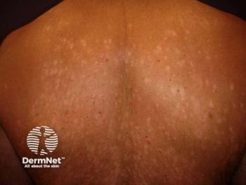
Comparative diagnosis of actinic keratosis (AK) and squamous cell carcinoma (SCC) in situ is needed for accuracy in treatment plan.
Advertisement
Making an accurate diagnosis of
While the definitions of actinic keratosis, SCC in situ and invasive SCC are well-defined and agreed upon, the interpretation of the histology can be very subjective and may differ among pathologists and their individual judgement of key histologic parameters.
Therefore, it is crucial for clinicians to include clinical information regarding the lesion for correlation. This will help a pathologist arrive at an accurate diagnosis, which enables the dermatologist to prescribe the most appropriate therapy, says Travis Vandergriff, M.D., associate professor in the department of dermatology, and dermatopathologist at UT Southwestern Medical Center, Dallas.
“There is a clear consensus regarding the definitions of these lesions but then making the judgement of whether or not some of the criteria that we use to distinguish them are actually fulfilled is a lot more subjective than I think people realize,” he notes.
The main difference between SCC in situ and AK is that in SCC in situ, the full thickness of the epidermis is involved with atypical proliferation of keratinocytes; whereas, in AK, the atypia is limited to lower levels of the epidermis and not its full thickness. The critical point is making the histopathologic judgement on whether the full thickness is involved with atypia or perhaps just the lower two-thirds of the epidermis. This judgement differs among individual pathologists and, according to Dr. Vandergriff, there is some discretion that pathologists use to decide whether or not the criteria are met.
Similar to the assessment of dysplastic nevi and other melanocytic neoplasms, there is a certain amount of interpretation that is involved by the pathologist and, as such, the inter-observer agreement can often diverge. Dr. Vandergriff stresses that this is not due to a lack of education or knowledge about the criteria, but more about the subjective interpretation of the individual pathologist if the criteria are being met.
“When the AK under scrutiny is meeting all the classic features like basal layer atypia and intermittent parakeratosis, you would generally have a really high level of agreement among pathologists. However, when it gets closer to SCC carcinoma in situ and the lines start to blur between our definition of AK and SCC in situ, that’s when there is more dispute among pathologists,” Dr. Vandergriff says.
When pathologists can incorporate clinical information into interpreting a case they can arrive at a more accurate diagnosis, Dr. Vandergriff says. For example, including detailed information not only regarding the lesion specifics but also about the patient, such as noting patients who are immunosuppressed or have significant sun damage and therefore may have a higher risk of developing SCC, are data points that may inform a pathologist if a lesion appears to be borderline.
“The clinical information about the patient and the lesion in question can be key in helping influence the pathologist’s interpretation of the lesion, directly impacting the therapeutic choices of the clinician,” Dr. Vandergriff says. “It is important to correlate with clinical information and to recognize that there are these borderline or in-between lesions, and that is where some discretion is needed and where clinical correlation becomes important.”
According to Dr. Vandergriff, the problem relates to the fact that clinicians and pathologists are trying to put labels on specific lesions and make them binary diagnoses when, in reality, these lesions are part of a spectrum, and they can fall anywhere along that spectrum. Trying to decide exactly where the lesion falls on the spectrum relies on the discretion of the individual pathologist, along with clinical correlation.
Most dermatologists and pathologists would agree that borderline lesions or advanced AK's or any lesion that is concerning for early SCC in situ development are lesions that definitely require some form of treatment. Surgery is not often the first choice, as other less invasive destructive therapies including
“We always try to convey in the pathological report if the lesion is advanced or borderline SCC in situ and, accordingly, the clinician can implement a measured and appropriate therapy,” Dr. Vandergriff says.
Articles in this issue
over 6 years ago
Non-melanoma skin cancer increases in the elderlyover 6 years ago
Looming medical billing and coding changes in healthcare policyalmost 7 years ago
Radiodermatitis increasingly common in female breast cancer patientsalmost 7 years ago
7 tips to get the most from your social media postsalmost 7 years ago
IL-17 inhibitors may fill treatment gap in rosaceaalmost 7 years ago
Head and neck melanoma characteristicsalmost 7 years ago
Do continuing medical education certifications improve patient safety?Advertisement
Related Content
Advertisement


Latest CME


Advertisement
Advertisement
Trending on Dermatology Times
1
GX-03 Phase 2 Program Expands to Full Atopic Dermatitis Spectrum
2
Teva Advances TEV-'408 Into Phase 2b for Vitiligo
3
Topical GX-03 Shows Early Promise in Mild to Moderate Atopic Dermatitis
4
First-in-Class Oral Piclidenoson Reaches Pivotal Phase 3 Enrollment Milestone in Plaque Psoriasis
5