MGH melanin research identifies pigmentation process
Boston - The question of how melanin - the pigment responsible for skin and hair color in mammals - is delivered to appropriate locations may have been answered in a study conducted by researchers at the Massachusetts General Hospital Cutaneous Biology Research Center, MedicalNewsToday.com reports.
Boston - The question of how melanin - the pigment responsible for skin and hair color in mammals - is delivered to appropriate locations may have been answered in a study conducted by researchers at the Massachusetts General Hospital Cutaneous Biology Research Center, MedicalNewsToday.com reports.
In humans, melanin is deposited in the skin and hair. In mammals such as mice, however, melanin is primarily deposited in the coat, leaving the skin beneath unpigmented. Melanocytes deposit melanin via cellular extensions called dendrites that reach out to other cells in the epidermis or the hair follicles. The focus of the study was the mechanism determining whether melanin is delivered to a particular cell, which has been unknown.
The MGH-CBRC researcher team theorized that a mouse gene known as Foxn1 could play a role. Lack of Foxn1 is responsible for hairless mice and other defects of the skin. A similar phenomenon exists in humans with inactivation of the corresponding gene.
When the researchers developed a strain of transgenic mice in which Foxn1 is misexpressed in cells that do not usually contain melanin, they found those normally colorless areas became pigmented. Examining the skin of the transgenic mice revealed that melanocytes were contacting and delivering melanin to the cells in which Foxn1 was abnormally activated. No pigment was observed in the corresponding tissues of normal mice.
When the researchers examined human skin samples, they found that the human version of Foxn1 was also expressed in cells known to be pigment recipients. Further experiments revealed that Foxn1 signals melanocytes through a protein called Fgf2, levels of which rise as Foxn1 expression increases.
The researchers note the Foxn1/Fgf2 pathway probably embodies other functions in the skin, and it is probably not the only pathway responsible for the targeting of pigment. They write that their next phase of research will be to identify other genes that can confer the pigment recipient phenotype or control the targeting of pigment.
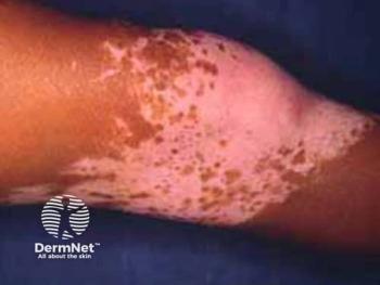
The results of the research may eventually be relevant to disorders such as vitiligo, age spots, the graying of hair and even melanoma.
Newsletter
Like what you’re reading? Subscribe to Dermatology Times for weekly updates on therapies, innovations, and real-world practice tips.


