

|Articles|March 25, 2022
Practical Management of Birthmarks and Hamartomas
Author(s)Keith Loria
In a presentation at the current AAD annual meeting, experts discussed differential diagnoses and clinical presentations in birthmarks and hamartomas.
Advertisement
This week, the American Academy of Dermatology (AAD) Association’s annual meeting returned live and in-person in Boston, Massachusetts. In the scientific seminar, “Practical Management of Birthmarks and Hamartomas,” 5 presenters discussed the practical management of a variety of birthmarks and birthmark-like conditions including hemangiomas, vascular stains, melanocytic nevi, nevus sebaceous, and hamartomas using a check-list approach.
The experts discussed diseases and clinical presentations posing questions such as “What should I be thinking about?” “Are any tests needed?” “Is there a window of opportunity to intervene?” “Do I (or others) need to see patients back in a follow-up appointments and if so what should we be looking for?” and “How do I counsel parents about what to expect over time?”
Renee M. Howard, MD, FAAD, pediatric dermatologist at University of California, San Francisco (UCSF) Benioff Children’s Hospital Oakland, California, presented a section entitled, “Developmental Hotspots: When to Worry, What to Do,” offering a checklist approach for diagnosis and treatment and outlining differential diagnosis of developmental anomalies with skin findings at 4 hotspots—the nose, brow, scalp, and lower back.
“My talk [covers] how to evaluate skin lesions that result from developmental mistakes that present in the skin, though some of them have deep connections. So you can’t approach them only as a dermatologist,” Howard says. “You have to think about what to image, where to refer, and specifically, I’m focusing on anomalies in high-risk locations.”
When Howard was a resident in dermatology in the early 1990s, she saw a patient with a midline cervical cleft, a rare anomaly on the anterior neck, and she couldn’t find much information about the condition. That led her to becoming interested in developmental birthmarks.
“It turns out that not just the front of the neck, but the front of the ear, nose, scalp, back, and also around the umbilicus are really high-risk areas for these skin findings,” Howard said.
She said it’s important to educate dermatologists about developmental birthmarks because they need to be managed differently and sometimes present later in adulthood.
“If you see a skin lesion that has a deep extension because it has a developmental origin—for example a cyst that’s on the middle of the lower back—that deep component can extend into the spinal cord,” Howard said. “As a dermatologist, you shouldn’t be managing that the way you would a regular cyst in the skin. It’s important to be knowledgeable about what lesions are high-risk.”
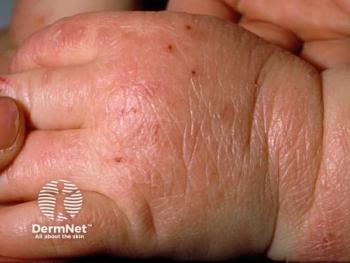
During her 25-minute presentation, Howard showed photos of patients that she and her colleagues have seen in their practices, with pits, nodular growths, and aplasia cutis congenita—the absence of skin in a focal area).
“I…talk about how to manage lesions in each of the 4 locations—nose, brow, scalp, and lower back—what causes them, how you image them, and when you need to refer,” she said.
For example, high risk and cutaneous signs for lumbosacral (LS) spine include midline mass, atypical dimple, thick tuft of terminal hair, membranous aplasia cutis congenita (ACC), tail or tag, and segmental hemangioma.
Other important signs to be discussed include a ring of dark hair, midline defects, and other high-risk features to look out for.
“I hope people will feel confident that they can recognize any skin lesions that may be overlying a developmental anomaly with deep extension and feel confident on how to manage that,” Howard said.
During another part of the program, pediatric dermatologist Ilona J. Frieden, MD, FAAD, director of UCSF Birthmarks & Vascular Anomalies Center, discusses the features of vascular birthmarks, particularly those which may have extra-cutaneous associations.
She discussed nevus simplex complex, and possible associations; what are considered regular port-wine stains, with potential concerns and approach; 3 types of special vascular stains; and other conditions in the differential diagnosis.
While previously, all vascular stains were lumped under port-wine stain or capillary malformation, further delineation can help guide management, she explained
Concerning nevus simplex, Frieden pointed out that it is the most common vascular birthmark present at birth, between 20% to 60%. While it occurs in all races and skin types, it may be easier to appreciate in those with lighter skin tones.
“Most cases are not associated with extra-cutaneous dz [disease],” she says. “However, there are someextracutaneous associations including megancephaly-capillary malformation (M-Cap), beckwith weidemann syndrome, a nevus-simplex like stain overlying developmental anomaly (eg, a lump, lipoma, hypertrichosis, aplasia cutis); Roberts SC [pseudo-thalidomide syndrome], and nova syndrome.”
Highlighing what are considered regular port-wine stains, Frieden explained how they virtually always present at birth and discussed those that may have added risks. For example, the risk of Sturge-Weber syndrome should be considered if the port-wine stains involved the forehead and size is an additional determinant or risk. In a recent study of 96 infants with forehead port-wine stains, Sturge-Weber syndrome was present in 22%, nearly all in those with larger port-wine stains.
The presentation also included exam essentials for major differential diagnoses, widespread blotchy stains with overgrowth and worrisome and not-so worrisome features, and a trio of special vascular stains—Cutis marmorata telangiectatica congenita (CMTC), geographic stains, and fast flow stains.
Frieden provided a checklist as to what to look out for, the care involved, and associated risks. She concluded her talk by discussing the differential diagnosis of vascular stains including infantile hemangioma precursors and tufted angiomas.
Howard noted in her session that if a case has a vascular stain that also has another skin finding in a high-risk area, such as extra hair on the scalp, it is high risk for having a deep extension.
Reference:
Frieden, IJ, Howard RM, Hawryluk EB, Oza VS, Shah SD. Practical management of birthmarks and hamartomas. Presented at: American Academy Dermatology Association 2022 Annual Meeting; March 25-29, 2022; Boston, MA.
Advertisement
Related Content
Advertisement


Latest CME


Advertisement
Advertisement
Trending on Dermatology Times
1
Switching from Biologics to Upadacitinib Yields Progressive Improvements in Moderate to Severe AD
2
Systemic JAK Inhibitors for Vitiligo: Emerging Clinical Trial Data and Safety Considerations
3
Real-World Study Finds Experienced Dermatologists Outperform AI in Skin Cancer Diagnosis
4
How Dermatologists Can Incorporate GLP-1 Receptor Agonists Into Psoriasis Care
5