
|Articles|March 25, 2021
- Dermatology Times, April 2021 (Vol. 42, No. 4)
- Volume 42
- Issue 4
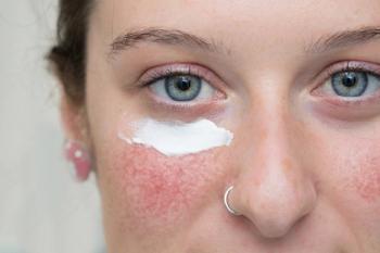
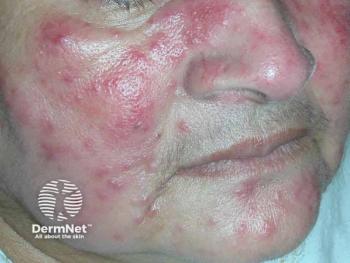
Collagen Degeneration Linked to Rosacea
Author(s)John Jesitus
Findings from a recent study indirectly support therapeutic approaches aimed at restoring the dermal matrix.
Advertisement
Recent study results linking photodamage-induced collagen degeneration with development of erythematotelangiectatic rosacea (ETR) suggest support for use of treatments aimed at improving the dermal collagen matrix.1
Led by Anna Chien, MD, investigators used a case-control approach to seek a scientific basis to explain the physical finding of telangiectasias in ETR, says coauthor Sewon Kang, MD, in an interview with Dermatology Times®. Translational techniques led investigators to focus on degraded collagen, a finding they were able to link to earlier observations that, in such situations, tubelike vascular structures can be made from endothelial cells.
Kang is chairman of the Department of Dermatology and the Noxell Professor of Dermatology at Johns Hopkins School of Medicine (JHSM) in Baltimore, Maryland. Chien is codirector of the Cutaneous Translational Research Program in the Department of Dermatology and an associate professor of dermatology at JHSM.
The present study included 5 patients with mild to moderate ETR and 5 matched controls. Using Picro Sirius red staining for collagen visualization and CD31 for blood vessels, investigators stained 3-mm punch biopsies of facial skin from patients with ETR and normal facial skin from matched controls. To facilitate standardized comparisons of samples between patients with ETR and healthy controls, investigators used the ImageJ color thresholding tool (an open-source Java image-processing program) to generate numeric data from histological images.
Analysis showed that patients with rosacea had significantly less collagen content than controls (16.812% ± 7.787% vs 19.603% ± 8.821%, respectively; P=.030) and significantly greater mean microvessel density (4.775 E-5 ± 1.493 E-5 µm-3 versus 2.559 E-5 ± 8.732 E-6 µm-3; P=.004).1 Additionally, participants with rosacea had significantly higher mean microvessel lumen area (491.710 ± 610.188 µm2) than controls (347.879 ± 539.642 µm2; P=.003).
“The fact that all 5 patients with rosacea had increased microvessel density versus their matched control patients makes sense because we studied ETR patients’ skin,” Kang adds. “Our approach demonstrated that quantitatively, there is more blood vessel lumen density in the patients’ skin.”
The fact that patients with rosacea also had decreased collagen versus their matched controls supports investigators’ underlying hypothesis that in the context of damaged collagen, fibroblasts make less procollagen. “The findings are consistent with previous study results showing that when you have degraded dermal matrix, the fibroblasts are not making as much procollagen as before.2 With the broken collagen, you tend to set up the environment in favor of more vessel tube-like formation,” the authors wrote.
Their findings are consistent with a previous study that evaluated histologic and molecular differences between ETR and telangiectatic photoaging (TP). In this study, facial biopsies from patients with ETR exhibited increased vasodilation compared to controls.3 Additionally, expression of matrix metalloproteinases (MMPs)-1, -3 and -9 was significantly higher in patients with ETR versus controls, and MMP-3 expression was also significantly elevated in patients with ETR versus those with TP. ETR tends to appear more prominently in areas of skin prone to atrophic photoaging resulting from ultraviolet (UV) exposure, says Kang, and some of the mechanisms involved in UV-induced photoaging may be involved in the pathophysiology of rosacea, especially in ETR.
The present study showed a link between decreased collagen content and increased microvessel size and density in ETR-affected skin that was not found in control subjects. “These structural changes to the dermal matrix may represent early aberrations involved in promoting the progression of rosacea,” the authors wrote.
“Regarding the clinical subtype of rosacea that is dominated by telangiectasias, we can say with corroborating data from our previous work2 that dermal matrix degradation to a significant degree can promote telangiectasia formation,” Kang adds. “We can explain at the clinical level why that happened.”
Indirectly, the study supports therapeutic approaches aimed at restoring the dermal matrix. “If the logic says that dermal matrix degradation sets up the milieu for endothelial cells to make little tube-like structures that clinically look like telangiectasias, then a way to improve that situation is to restore the degraded dermal matrix [to] a healthier state,” Kang notes, adding that studies show that medical therapies such as topical retinoids and devices such as fractional lasers can improve the dermal matrix in photoaged skin.4,5
Limitations of the present study included its small size and limited scope (addressing only the ETR subtype). The use of immunohistochemical staining on tissue sections allowed investigators to identify the presence of degraded collagen, says Kang, but not to quantify it, as a hydroxyproline assay would.
However, he concludes, “We identified a correlation between decreased collagen content and increased microvessel size and density in rosacea patients that was not observed in controls. These structural changes to the dermal matrix may contribute to the characteristic vessel growth and dilation in rosacea.”
Disclosure:
Kang reported no relevant or financial interests.
References:
1. Thompson KG, Rainer BM, Leung S, Qi J, Kang S, Chien AL. The association of photo-induced collagen degeneration and the development of telangiectasias in rosacea. J Anat. Published online January 11, 2021. doi:10.1111/joa.13394
2. Varani J, Perone P, Warner RL, et al. Vascular tube formation on matrix metalloproteinase-1-damaged collagen. Br J Cancer. 2008;98(10):1646-1652. doi:10.1038/sj.bjc.6604357
3. Helfrich YR, Maier LE, Cui Y, et al. Clinical, histologic, and molecular analysis of differences between erythematotelangiectatic rosacea and telangiectatic photoaging. JAMA Dermatol. 2015;151(8):825-836. doi:10.1001/jamadermatol.2014.4728
4. Kang S. The mechanism of action of topical retinoids. Cutis. 2005;75(suppl 2):10-13; discussion 13.
5. Borges J, Araújo L, Cuzzi T, Martinez L, Gonzales Y, Manela-Azulay M. Fractional laser resurfacing treats photoaging by promoting neocollegenesis and cutaneous edema. J Clin Aesthet Dermatol. 2020;13(1):22-27.
Articles in this issue
about 5 years ago
Regenerative Medicine Gaining Popularityabout 5 years ago
Combination Foam May Benefit Patients of Colorabout 5 years ago
AI Diagnostics Fall Short in Skin of Colorabout 5 years ago
Microneedling Strategiesabout 5 years ago
Neuromodulators Combat Key Rosacea Challengesabout 5 years ago
Baricitinib Demonstrates Positive Outcomes for Alopecia Areataabout 5 years ago
COVID-19 Vaccine Refusal: Grounds for Termination?about 5 years ago
Tazarotene Excels Across BMI Categoriesabout 5 years ago
Solving Pierced Earring Challengesabout 5 years ago
Emerging Agents Augment Melasma ModalitiesAdvertisement
Related Content
Advertisement


Latest CME


Advertisement
Advertisement
Trending on Dermatology Times
1
Are Private Dermatology Practices at Risk for Extinction? A New Survey Sheds Light
2
Upadacitinib Receives Positive CHMP Opinion for Severe Alopecia Areata
3
Dermatology Times June 2026 Print Recap
4
Why Joint Damage Inhibition Matters in Psoriatic Arthritis Treatment
5