
|Articles|May 1, 2005
West Nile virus skin symptoms documented
New Orleans — Scientific literature rarely portrays cutaneous manifestations of West Nile virus (WNV) because these manifestations are, by nature, transient. However, a recent case report contains what is only the second publication of these symptoms.
Advertisement
"It's not unusual to have a cutaneous eruption with WNV," says Padma Nallamothu, M.D., chief resident, department of dermatology, Henry Ford Health System, Detroit. "It has not been reported in terms of the clinical pictures and histology because it tends to be fleeting. Furthermore, reports of WNV infection usually report a 'rash' without describing it or telling us what to look for."
Diagnostic clues In September 2002, a 41-year-old woman presented to an emergency room shortly after returning from a camping trip in northern Michigan.
The patient was admitted to the health system's infectious diseases service, and the dermatology service was consulted primarily for assistance with the patient's cutaneous eruption, which occurred in conjunction with the above symptoms.
"Her eruption looked more like a viral exanthem," Dr. Nallamothu says. "These skin findings, together with her travel history and systemic symptoms, led us to check WNV IgM levels. Meningitis was ruled out based on a negative lumbar puncture, and instead of empirically treating her with IV antibiotics, we were able to say, 'this looks more like a viral eruption more consistent with WNV.' "
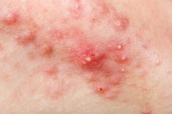
Physical examination of the patient's skin revealed a diffuse monomorphic eruption comprised of multiple erythematous papules measuring 1 mm to 2 mm. The eruption spared the patient's face, palms and soles.
Histological abnormality Besides the positive serum WNV IgM, the only histological abnormality doctors noted was a decreased white blood cell count.
The patient's cerebrospinal fluid showed no signs of meningitis, while viral cultures for enterovirus, Coxsackie virus and echovirus proved negative, as did tissue bacterial cultures. The patient had normal chemistry and liver profiles, nonreactive RPR, negative blood cultures and negative ANA. Mono Ab and RF screens also were normal. A head CAT scan also was negative.
Treatment With treatments including topical triamcinolone acetonide cream 0.1 percent and oral hydroxyzine, the eruption resolved in about a week, which is typical for cutaneous WNV manifestations.
"The skin findings usually present early on in the course of the infection and have been described as fleeting," Dr. Nallamothu says. "Often, patients don't present to a physician because the symptoms are mild and short-lived."
Diagnosing WNV infections may be difficult because most dermatologists' experience with WNV is minimal at best.
"The infection has only been present in the United States since 1999," Dr. Nallamothu notes. "And patients frequently present because of neurologic symptoms, not cutaneous symptoms."
That is, if they present at all. Neurologic symptoms afflict approximately 50 percent of West Nile patients. Skin symptoms occur in about 20 percent.
"It looks like this eruption is fairly classic in the condition. But it's not classic to the condition (Anderson RC et al. J Am Acad Dermatol. 2004 Nov;51(5):820-823)," she says.
Differential diagnoses for similar-looking eruptions include Rocky Mountain spotted fever and other viral infections.
Newsletter
Like what you’re reading? Subscribe to Dermatology Times for weekly updates on therapies, innovations, and real-world practice tips.
Advertisement
Related Content
Advertisement


Latest CME


Advertisement
Advertisement
Trending on Dermatology Times
1
AbbVie Files for Vitiligo Indication, Putting Systemic Therapy Under Regulatory Review
2
Nutrafol Expands Portfolio with First and Only Hair Loss Supplement for Male Patients 50 and Older
3
Introducing Dermatology Times NP/PA Connect
4
Narrow-Spectrum Sarecycline Approved for Moderate to Severe Acne in China
5