

|Articles|May 11, 2022
Treating Seborrheic Dermatitis
Author(s)Sherry G. Cohen, NP-C
A look at the common condition and how to treat it.
Advertisement
A healthy, 6-week-old boy was evaluated for a 2-week history of asymptomatic progressive thick scale and mild erythema over his scalp, extending to his forehead. His examination was otherwise normal.
What’s the diagnosis?
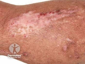
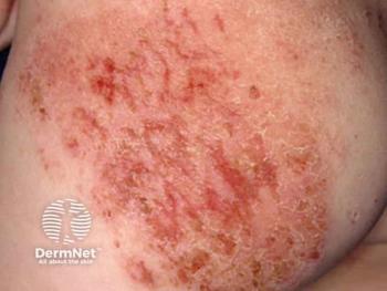
The diagnosis is seborrheic dermatitis, sometimes called cradle cap when it appears on the scalp. This chronic inflammatory skin condition is present worldwide and affects all age groups and sexes equally. It is a common disorder, and the diagnosis is usually made clinically.
Infants and young children tend to have mild disease that lasts 4 to 6 weeks but can persist up to a year. In infants, it presents with erythema and thick, patchy, white or yellow scales, sometimes with crust on the scalp and intertriginous areas including the axilla, retroauricular creases, and diaper area, in the first few months of life . On the face, it appears on the central and lower forehead, eyelids, cheeks, and nasolabial folds.1
Seborrheic dermatitis also tends to occur in adolescents, affecting 2% to 5% of that population and appearing as an itchy, scaly rash on the scalp, face, presternal area, and skinfolds.2 On darker-pigmented children, postinflammatory hypopigmentation may persist for several weeks to months after clearing with treatment.3
The cause of seborrheic dermatitis is not known but has been attributed to androgens that pass from mother to baby during pregnancy. In newborns and infants, androgens stimulate the growth of sebaceous glands that might drive the process. The yeast Malassezia can be found in higher concentrations on patients who have seborrheic dermatitis compared with controls and who may have an inflammatory reaction.2
The differential diagnosis includes psoriasis, atopic dermatitis, contact dermatitis, Langerhans cell histiocytosis, and acrodermatitis enteropathica or nutritional deficiency.2,4
In young children, psoriasis usually starts in the diaper area and is well delineated. In infants, seborrheic dermatitis and atopic dermatitis can look very similar. The best way to distinguish them is by location and the presence of intense itching in atopy. Both conditions can cause a red, scaly scalp. The rash of seborrheic dermatitis generally involves the skinfolds and diaper area, which are usually spared in atopic dermatitis. Atopic dermatitis often appears on the face, shins, forearms, and other exposed areas that infants can scratch. Contact dermatitis is often seen on the face or diaper area and is itchy. Langerhans cell histiocytosis presents with persistent hemorrhagic, crusted, atrophic, and/or scaly patches that can be anywhere but tend to appear in the skin creases.
Infantile seborrheic dermatitis often resolves on its own in a few months. Treatment options for persistent and/or symptomatic lesions include low-potency topical steroids (eg, hydrocortisone 1% ointment); topical calcineurin inhibitors (eg, pimecrolimus cream for > 3 months of age, tacrolimus ointment for > 2 years of age); topical antifungal creams, ointments, or shampoos (eg, ketoconazole, selenium sulfide); and mild keratolytic agents (eg, urea 10%-20%). Oil preparations such as mineral oil can soften and loosen the scale, making it easier to separate from the hair and scalp. For areas that are not as sensitive as the face or intertriginous areas, a higher-potency topical steroid can be used sparingly for a short course; antifungals can be relied on for long-term therapy. Other medicated shampoos and treatments include ingredients such as zinc pyrithione, coal tar, and ciclopirox olamine. For severe cases, oral antifungals are sometimes used.5,6
The findings of 1 review of multiple, randomized, controlled trials showed that ketoconazole, ciclopirox olamine, and steroids had similar effective results, but the antifungals showed fewer adverse effects. Moreover, the antifungals showed comparable efficacy.7
For this 6-week-old patient, the parents were instructed to massage the infant’s scalp with mineral oil to loosen the scale before washing with ketoconazole shampoo daily. Over the following 2 weeks, the rash resolved, and the infant subsequently remained clear.
References
1. Tom WL, Eichenfield LF. Eczematous disorders. In: Eichenfield LF, Frieden IJ, Mathes EF, Zaenglein AL, eds. Neonatal and Infant Dermatology. Elsevier; 2015:225-226.
2. Hogan PA, Langley RGB. Papulosquamous diseases: seborrheic dermatitis. In: Schachner LA, Hansen RC, eds. Pediatric Dermatology. Elsevier; 2003:924-927.
3. Elgash M, Dlova N, Ogunleye T, Taylor SC. Seborrheic dermatitis in skin of color: clinical considerations. J Drugs Dermatol. 2019;18(1):24-27.
4. Galimberti F, Mesinkovska NA. Skin findings associated with nutritional deficiencies. Cleve Clin J Med. 2016;83(10):731-739. doi:10.3949/ccjm.83a.15061
5. Okokon EO, Verbeek JH, Ruotsalainen JH, Ojo OA, Bakhoya VN. Topical antifungals for seborrhoeic dermatitis. Cochrane Database Syst Rev. 2015;(5):CD008138. doi:10.1002/14651858.CD008138.pub3
6. Kastarinen H, Oksanen T, Okokon EO, et al. Topical anti-inflammatory agents for seborrhoeic dermatitis of the face or scalp. Cochrane Database Sys Rev. 2014;(5):CD009446. doi:10.1002/14651858.CD009446.pub2
7. Clark GW, Pope SM, Jaboori KA. Diagnosis and treatment of seborrheic dermatitis. Am Fam Physician. 2015;91(3):185-190.
Advertisement
Related Content
Advertisement


Latest CME


Advertisement
Advertisement
Trending on Dermatology Times
1
FDA Accepts Addition of Bemotrizinol as First New Sunscreen Ingredient in 20 Years
2
AI Analysis Reveals Menopausal Hair Loss Linked to Systemic Health and Inflammation
3
Dual-Action Microneedle Patch Shows Promise for Psoriasis in Preclinical Study
4
Novel Botanical Moisturizer Demonstrates Superior Efficacy Over Metronidazole in Phase 2 Rosacea Trial
5