

|Articles|December 10, 2021
Treating and Managing Seborrheic Dermatitis
Author(s)Rachael Zimlich, RN, BSN
Seborrheic dermatitis will affect roughly half of all infants, making knowing how to treat and manage it key.
Advertisement
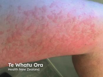
Cradle cap—or seborrheic dermatitis (SD)—is a common dermatologic condition that affects nearly half of all infants at some point in the first few months of their lives.1
This article will explore how to recognize and diagnose SD, as well as tips for treating and managing this condition.
What is SD?
SD is a chronic inflammatory skin condition sometimes referred to as cradle cap when it appears on the heads of infants, said Sherry G Cohen, NP-C, a family nurse dermatology practitioner in Lutherville, Maryland. It’s a common disease that is usually diagnosed clinically, and can present with mild disease lasting 4 to 6 weeks on average, but can last as long as a year.2
SD and dandruff fall into the same group of diseases that cause dry, flaky skin in the seborrheic areas of the body. Although dandruff is limited to the scalp alone, SD can affect other areas like the face, ears, nose, and eyelids.
This condition is most common during 3 stages of life1:
- The first 3 months of infancy
- During puberty
- Between the ages of 40 and 60 years
The scalp is most commonly affected in infants, earning this condition the name of cradle cap, but the face and diaper areas can also show signs of SD. It’s also important to know how it may appear differently on other parts of the body.1
- Scalp: Red to yellow plaques coated by thick, greasy scales
- Face and neck: Red or pink flaky plaques that can appear on the forehead, eyebrows, eyelids, nasolabial folds, and retro-auricular areas
- Body folds: Moist, shiny but non-scaly lesions common in the neck, armpits, and groin areas
- Trunk: Redness and scaling as limited plaques throughout the lower abdomen
Generalized SD is rare and is often a sign of other conditions such as Leiner’s disease or immunodeficiencies. In these cases, itching is mild or absent with widespread redness, and other symptoms like diarrhea or failure to thrive may be evident.1
Only about 2% to 5% of teenagers experience SD, and by this age it tends to appear as an itchy, scaly rash on the scalp, face, chest, and skin folds. Although SD affects different genders and ethnicities equally, darker-skinner children may have areas of more pronounced hypopigmentation that lasts for weeks after an area of SD has cleared.3
The exact cause of SD is unknown, Cohen said, but androgens passed from mother to baby during pregnancy are believed to play a role.
“In newborns and infants, androgens stimulate the growth of the child's sebaceous glands that might drive the process,” Cohen stated, adding that a yeast called Malassezia has been found to grow in higher concentrations on people with SD, possibly as a result of an inflammatory reaction to the yeast.
Diagnosing SD
Making a clinical diagnosis of SD takes a few steps, particularly in ruling out other possible causes for dermatitis.
Differential diagnoses for SD can include things like:
- Psoriasis
- Contact dermatitis
- Langerhan cell histiocytosis
- Acrodermatitis enteropathica
- Nutritional deficiency
- Atopic dermatitis
- Tinea capitis
- Rosacea
- Systemic lupus erythematous
In young children, a diagnosis can be narrowed based on how the dermatitis developed, Cohen added.
“Psoriasis usually starts in the diaper area and is well delineated. In infants, seborrheic dermatitis and atopic dermatitis can look very similar. The best way to distinguish them is by location, and the presence of intense itching in atopy,” Cohen explained. “Both can be red and scaly on the scalp. The rash of seborrheic dermatitis usually involves the skin folds and diaper area which are usually spared in atopic dermatitis. In infants atopic dermatitis often appears on the face, shins and forearms and other exposed areas that they can scratch. Contact dermatitis is often seen on the face or diaper area and is also itchy.”
A history and physical are the primary tools of diagnosis for SD, although skin biopsies may be performed in rare or complicated cases.
Management and treatment of SD
Infantile SD often resolves without treatment in a few months, Cohen said, but treatment options for persistent or symptomatic lesions can include medications like:
- Low potency topical steroids such as hydrocortisone 1 % ointment
- Topical calcineurin inhibitors like pimecrolimus cream or tacrolimus ointment depending on age
- Topical antifungal creams, ointments, or shampoos like ketoconazole or selenium sulfide
Other remedies that may help to soothe the affected areas include oil preparations like mineral oil. This can soften and loosen the scales caused by SD, making it easier to separate the hair from the scalp, Cohen stated.
For areas that are not as sensitive as the face or intertriginous areas, a higher potency topical steroid can be used sparingly for short courses, whereas antifungals are preferred for long-term therapy, she added, explaining that studies have shown similar efficacy between these 2 treatments with lower adverse effects in the antifungals.4,5,6,7
Other medicated shampoos and treatments that might be used include zinc pyrithione, coal tar, and ciclopirox olamine. Rarely, in the most severe cases, antifungals are given orally for systemic effect, Cohen said.
A referral to a pediatric dermatologist may be appropriate in cases of SD that are complicated, prolonged, or appear with other symptoms.
References
1.Borda LJ, Wikramanayake TC. Seborrheic dermatitis and dandruff: a comprehensive review. J Clin Investig Dermatol. 2015;3(2):10.13188/2373-1044.1000019. doi:10.13188/2373-1044.1000019
2.Eichenfield LF, Tom WL, Chamlin SL, et al. Guidelines of care for the management of atopic dermatitis: section 1.diagnosis and assessment of atopic dermatitis. J Am Acad Dermatol. 2014;70(2):338-351. doi:10.1016/j.jaad.2013.10.010
3. Elgash M, Dlova N, Ogunleye T, Taylor SC. Seborrheic dermatitis in skin of color: clinical considerations. J Drugs Dermatol. 2019 Jan 1;18(1):24-27.
4. Galimberti F, Mesinkovska NA. Skin findings associated with nutritional deficiencies. Cleve Clin J Med. 2016;83(10):731-739. doi: 10.3949/ccjm.83a.15061.
5. Okokon EO, Verbeek JH, Ruotsalainen JH, Ojo OA, Bakhoya VN. Topical antifungals for seborrhoeic dermatitis. Cochrane Database Syst Rev. 2015;(5):CD008138. doi:10.1002/14651858.CD008138.pub3
6. Kastarinen H, Oksanen T, Okokon EO, et al. Topical anti-inflammatory agents for seborrhoeic dermatitis of the face or scalp. Cochrane Database Syst Rev. 2014 19;2014(5):CD009446. doi: 10.1002/14651858.CD009446.pub2.
7. Clark GW, Pope SM, Jaboori KA. Diagnosis and treatment of seborrheic dermatitis. Am Fam Physician. 2015;91(3):185-90.
Advertisement
Related Content
Advertisement

Latest CME


Advertisement
Advertisement
Trending on Dermatology Times
1
Once-Daily Zasocitinib Offers New Oral Option for Psoriasis
2
MoonLake Reports Positive 52-Week Phase 3 Data for Sonelokimab in Hidradenitis Suppurativa
3
Top 5 Articles of the Month: June 2026
4
Combination Therapy Guides Acne Management Across Severity Levels
5