The merger, backed by an oversubscribed $180 million private placement, will fund development of NOR-101, an IL-13 x IL-18 bispecific antibody entering phase 1 trials in atopic dermatitis in early 2027.
According to new research, AI reads real-world smartphone photos to score 4 eczema severity signs, boosting EASI training accuracy and reducing rater variability.
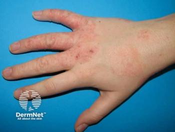
Accurate assessment of atopic dermatitis (AD) severity is important for guiding treatment and monitoring outcomes, but as noted in prior research, the Eczema Area and Severity Index (EASI) relies on subjective visual scoring that can contribute to inter-rater variability.1 A prospective, multicenter study evaluated whether convolutional neural network (CNN)-based artificial intelligence (AI) software could assess 4 EASI signs from smartphone images captured without cropping, masking, or controlled imaging conditions.2
The study included 1016 adults with AD enrolled across 16 institutions in Japan between October 2023 and March 2024. Participants provided 3676 skin images captured using a mobile smartphone. Investigators photographed representative lesions from the head and neck, trunk, upper limbs, and lower limbs, with a maximum of four images per patient. Images were acquired under variable lighting and viewing angles and were not cropped or masked for analysis.
The AI model used the ConvNeXt architecture to predict continuous severity scores for 4 EASI signs: erythema, papulation/edema, excoriation, and lichenification. The model was trained using consensus severity grades from a Central Assessment Committee of board-certified dermatologists. Its performance was evaluated at EASI component severity thresholds of ≥1, ≥2, and 3 using receiver operating characteristic analysis.
For the study's primary endpoint, identifying an EASI component score of ≥2, the AI demonstrated moderate discriminative performance across all 4 signs. Area under the receiver operating characteristic curve (AUC) values ranged from 0.825 to 0.860. Erythema had the highest accuracy, with an AUC of 0.860, while papulation/edema had the lowest, at 0.825. Sensitivities ranged from 0.745 to 0.873, and specificities ranged from 0.675 to 0.764.
Performance was stronger when identifying the most severe component score of 3. AUC values exceeded 0.900 for all 4 signs, ranging from 0.901 to 0.959. Excoriation demonstrated the highest accuracy and reached a sensitivity of 1.000, while erythema achieved the highest specificity, at 0.906. The authors suggested that more severe lesions may be easier for the AI to recognize because their clinical features are more visible and distinct in photographs.
The model also maintained AUC values above 0.700 when identifying signs with a component score of ≥1, with values ranging from 0.778 to 0.832. The lower performance at this threshold may reflect the difficulty of distinguishing mild disease from near-normal skin. The study excluded patients with a total EASI score of 0, so the dataset did not include completely lesion-free skin.
Differences in performance among the 4 signs were also observed. Erythema performed best, potentially because its color changes are readily captured in standard photographs. Excoriation also showed relatively strong performance, whereas papulation/edema was more difficult to assess, potentially because these lesions are raised and three-dimensional but were evaluated using two-dimensional images.
The authors emphasized that the AI software was developed as an educational tool rather than for direct clinical decision-making. Its ability to evaluate uncropped and unmasked smartphone images could support standardized EASI training and potentially reduce variability among raters. However, further validation is needed before clinical applications can be considered.
Despite its promise, several limitations remain. The dataset consisted primarily of Japanese patients, leaving the model's performance in other populations and skin phototypes uncertain. The model did not estimate affected body surface area and therefore could not calculate a total EASI score. In addition, AD diagnoses were made according to routine clinical practice at individual sites without a single standardized diagnostic criterion.
Overall, the findings suggest that ConvNeXt-based AI software can assess individual EASI signs from real-world smartphone images with moderate-to-high accuracy, although broader validation is needed.
“With its low-cost and scalable design, this AI tool may offer a practical solution for training health care professionals and promoting standardized, efficient education in EASI severity assessment,” the authors concluded.1
References
1. Jacobson ME, Morimoto RY, Leshem YA, et al. The Eczema Area and Severity Index: An update of progress and challenges in its measurement of atopic dermatitis after 20 years of use. J Eur Acad Dermatol Venereol. 2025;39(1):70-85. doi:10.1111/jdv.20248
2. Yamanaka K, Pak Y, Inda Y, et al. Educational Artificial Intelligence Software to Support Assessment of Atopic Dermatitis Severity. J Dermatol. Published online July 30, 2026. doi:10.1111/1346-8138.70434