- Dermatology Times, December 2018 (Vol. 39, No. 12)
- Volume 39
- Issue 12
A review of treatment for dermatofibrosarcoma
Dermatofibrosarcoma is a common, but unusual locally aggressive cutaneous tumor. Its characteristic tentacle can grow into surrounding fat, muscle and even bone. For dermatologists this means that understanding treatment options is a priority, says a physician reporting from EADV 2018.
PARISâDermatofibrosarcoma is a common, but unusual locally aggressive cutaneous tumor. Its characteristic tentacle can grow into surrounding fat, muscle and even bone. For dermatologists, such as Onofre Sanmartin, M.D., this means that understanding treatment options is a priority.
During the European Academy of Dermatology and Venereology (EADV) Congress in Paris last week, Dr. Sanmartin, of the Instituto Valenciano de OncologÃa in Spain, presented an overview the diagnosis and treatment for dermatofibrosarcoma.
DERMATOFIBROSARCOMA OVERVIEW
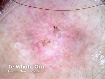
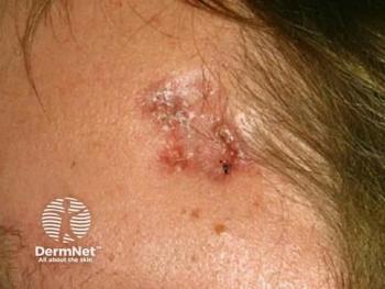
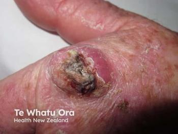
Dermatofibrosarcoma is most commonly found on the trunk, followed by proximal extremities and the head and neck. In the protuberans form (DFSP), the clinical presentation is plaques with some nodules. The non-protuberans form is more commonly seen in children.
Pathologically, dermatofibrosarcoma is easy to diagnose. It infiltrates the epidermis and becomes subcutaneous. It has a typical fibroblast-like proliferation with little or no mitosis, and the main characteristic is the tendency to spread into subcutaneous tissue with tumor tentacle-like extensions. Tumors can even reach the fascia and muscle in one third of patients.
Despite the characteristic histology, there are more than 10 clinical subtypes of DFSP and many different clinical presentations. Variants include fibrosarcomatous dermatofibrosarcoma, which has a fascicle appearance with a herringbone pattern. Another variety is subcutaneous, which is mainly found on the head and neck and characterized by no connection with the dermis or epidermis.
Immunohistochemical diagnosis is possible with 90-95 percent of patients positive for CD34 and 10 percent negative for CD34. Cytogenetics is the main tool for diagnosis, but DFSP is characterized by the translocation T (17;22)(q22;q13), which results in the platelet-derived growth factor B (PDGFB)/COL1A1 fusion gene. This causes overproduction of PDGFB, leading to PDFG receptor activation.
RNA gene sequencing (e.g. RT-PCR) can be used for diagnosis, as can FISH. “FISH is faster and sensitive enough, and should be recommended as a routine diagnostic tool,” Dr. Sanmartin said. Some patients (4 percent) with typical DFSP are negative for translocation in routine molecular screening, and 50 percent of cases defined as negative by FISH are positive with RT-PCR. While MRI is not useful for the lateral delineation of tumor or detection of a residual tumor, it can be suitable for large tumors and gauging the involvement of deep structures.
The main treatment for DFSP is surgery. The tumor must be completely removed. “This can be a challenge given its infiltrative nature,” said Sanmartin. The tumor extensions can be more than 5 cm away from the clinical part of the tumor, which means that much normal skin must be eliminated. DFSP also tends to recur in the same location. In one review, 2069 patients showed a recurrence rate of around 17 percent after surgery. It is possible that this standard surgery is not the best approach, Dr. Sanmartin said.
Only 0.84 percent recurrence is reportedly seen after Mohs micrographic surgery. This option can spare 50 percent of the skin at 2 cm, and according to Dr. Sanmartin should be used as the treatment of choice.
PDGF receptor inhibitors are an option for locally advanced and metastatic DFSP. Improved indication has been seen with imatinib mesylate, for example. However, Dr. Sanmartin considers 800 mg to be a very high dose of tyrosine kinase inhibitor (as has been used in previous studies) â he treats with 400 mg.
REFERENCE
Sanmartin, O. (2018). Dermatofibrosarcoma: From surgery to systemic therapy, The 27th European Academy of Dermatology and Venereology Congress, Paris, France, 15th September, 15:20 - 15:40.
Articles in this issue
about 7 years ago
Presentation and management of the neuropathic itchabout 7 years ago
Inadequately controlled atopic dermatitisabout 7 years ago
European data protection regulation impacts U.S. patient privacyabout 7 years ago
Market competition impacts topical generic medication pricingabout 7 years ago
High efficacy shown in self-inject psoriasis biologicabout 7 years ago
First approved biologic in dermatology to carry suffixabout 7 years ago
Steps to mitigate cyber riskabout 7 years ago
Dr. Reddy’s sells Cloderm to EPI Healthabout 7 years ago
ODAC returns to OrlandoNewsletter
Like what you’re reading? Subscribe to Dermatology Times for weekly updates on therapies, innovations, and real-world practice tips.

