

|Articles|October 14, 2022
- Dermatology Times, October 2022 (Vol. 43. No. 10)
- Volume 43
- Issue 10
Pediatric Nail Disorders
Author(s)Fazila Rajab
Management differs from adults and requires a meticulous examination to rule out underlying diseases.
Advertisement
Nail disorders among infants and children are a relatively uncommon observance in pediatric dermatology, but when they do appear, they can be associated with a systemic condition or a cosmetic or psychological problem.
Their prevalence varies among different populations. A study published in Turkey has found the incidence rate of nail disorders to be 4.4% in pediatric patients.1 Their management strategy differs from the strategy for adults and requires a thorough examination to rule out any underlying diseases.
Understanding the normal nail anatomy is vital for understanding nail disease. The nail plate, a keratinized structure, is attached to the vascularized nail bed at the hyponychium, paronychium, eponychium, and cuticle. The cuticle prevents foreign objects from entering the nail matrix, an epithelial structure beneath the nail. The lunula is the visible distal part of the nail matrix.
What follows are several nail conditions that may be seen in a pediatric dermatology practice.
Koilonychia
Koilonychia, commonly known as spoon nails, causes the nails to become abnormally concave. When a nail is in this state, it might look like it has been scooped out because of its pliable nature. The condition is prevalent among 33% of neonates and usually goes away on its own within the first decade of life when the nail plate strengthens.2
If it does not go away, the culprits of koilonychia are usually injury, inflammatory dermatosis, or iron deficiency anemia. Most cases in young toddlers are brought on by trauma caused by improperly fitting shoes or excessive thumb sucking. Dermatologists usually diagnose the condition by placing a drop of water on the nail plate. Treatment mostly depends on managing the underlying cause. Foods rich in iron such as beans and lentils, dark chocolate, fish, and spinach are the mainstay of treatment for koilonychia in children.3 Moreover, koilonychia can be an important predictor of underlying systemic conditions, including trichothiodystrophy, Darier disease, and LEOPARD syndrome.4
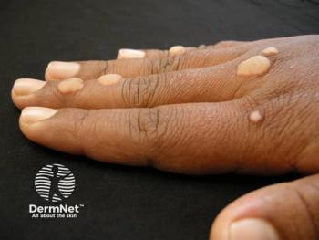
Onychocryptosis
Onychocryptosis, most commonly known as in-grown toenails, is among the most frequently seen nail condition. It may cause extreme discomfort and hamper a child’s everyday activities.5
Onychocryptosis is characterized by redness and swelling surrounding the nail folds of the toes. Advanced disease may include symptoms like seropurulent discharge, granulation tissue, and tissue hypertrophy. Affected individuals may experience trouble walking or be rendered immobile due to their pain intensity. Erythema, inflammation, or discharge around the nail edge are other potential complaints from patients.
Although the etiology of this condition is not clear, genetic susceptibility, trauma, tight-fitted shoes, and incorrectly trimmed toenails are some of the contributing factors. Other factors may include fungal infection, hyperhidrosis, and onychotillomania. Treatment is indicated in severe cases when the pain is affecting children’s daily activities. Patients are advised to reduce the risk of their children developing the condition by following tips such as having them wear comfortable shoes, treating underlying conditions, and asking for help trimming toenails.
Treatment can either be operative or nonoperative, depending on the patient’s condition. With nonoperative treatment, the patient’s toe is soaked in warm water for 10 minutes, followed by the application of antibiotic ointment twice a day. Operative options are indicated for patients who don’t respond to nonoperative treatment and may include complete or partial nail avulsion alone or in association with germinal matrix ablation.6
Trachyonychia
Trachyonychia presents with longitudinally ridged nails, commonly known as opaque trachyonychia, or may manifest in a uniform, opalescent pattern with pits, known as shiny trachyonychia.7
In most cases, a diagnosis of trachyonychia may be made based only on the patient’s clinical presentation. However, histological examination of nail clipping to detect fungal infection is a legitimate element of examining onychodystrophy. The matrix of nail unit samples taken for diagnosing trachyonychia typically displays a spongiotic pattern. A nail matrix specimen can be examined under a microscope to reveal histopathologic signs of the underlying disease, such as lichen planus or psoriasis, in cases of trachyonychia.
Improvements in trachyonychia have been recorded following intralesional steroid injections at doses such as 2.5 to 3 mg/mL and 10 mg/mL. In children, it is advisable to start with a lesser concentration to prevent any localized adverse effects. Other topical treatments include topical steroids, tazarotene (Tazorac) 0.1% gel, topical psoralen ultraviolet A (PUVA), and urea cream. Oral biotin and pulse-dosage oral steroids, such as 4 mg of betamethasone and low-dose acitretin (Soriatane), are some of the other effective treatments.8
Onychomycosis
Onychomycosis is an infection caused by a fungus that invades the nail bed. The affected nail may have a thick, yellow appearance and become coarse or brittle. Onychomycosis tends to run in families; however, research suggests that it is more common in children with Down syndrome and immunodeficiencies and may pose a severe health problem in such patients.9
Fungal nail infections may be diagnosed with a potassium hydroxide test, a fungal culture, or a histopathologic study of the nail plates. Dermoscopy, reflectance confocal microscopy, and artificial intelligence are just a few examples of the newer diagnostic tools that have been developed to enhance onychomycosis detection in the clinic, whereas mycological tools like molecular assays have been developed to better detect the fungus in the laboratory.
Because the toenail grows so slowly, therapy can be difficult and a full recovery could take a year or more. Systemic antifungal medicines like itraconazole (Sporanox), terbinafine (Lamisil), and fluconazole (Diflucan) and topical antifungal drugs like efinaconazole (Jublia), ciclopirox, and tavaborole (Kerydin) are typically used in therapy. Children’s nails are thinner and grow more quickly than those of adults, so they often react better to topical antibiotics.10
Beau Lines
Beau lines result from the decreased mitotic activity of keratinocytes in the proximal nail matrix, thinning the nail plate and causing transverse groove formation. The depth and width of the depression signify the degree of damage and duration of the insult, respectively. Damage, infection, or problems in the nail fold are all possible causes. Some other causative factors may include diabetes, hypocalcemia, and chemotherapeutic drugs. Post-natal onset is the norm; nevertheless, Beau lines may be observed in older children following a high fever or may be associated with zinc deficiency in rare cases. Beau lines in more than one nail may signify a systemic sickness, long-term exposure to a potentially harmful environment, or a chronic condition,11 and thicker lines might be due to long-term sickness or trauma exposure.12
The dermatologist might diagnose Beau lines after thoroughly examining the nails and reviewing the patient’s medical history. The thickness and number of lines may also indicate the cause. Unfortunately, there is no treatment for Beau lines, but treating the underlying cause may help improve the outcome.
Onycholysis
Onycholysis is a condition in which the nail bed separates from the nail plate, resulting in an opaque white appearance of the affected part. It is mainly caused by trauma, medications, or nail infections but is sometimes associated with autoimmune diseases.13 The condition may predispose to secondary nail infections, most commonly by Pseudomonas aeruginosa and Candida albicans.
Clinically, onycholysis can be diagnosed by taking a patient’s history and doing a physical exam to determine the root cause. If the root of the problem is unclear, an examination may be necessary. Often, bacteria and fungi are cultured from nail clippings or scrapings. Systemic causes of nail disease may necessitate further testing like a thyroid function test.
Treatment may vary with the underlying cause and require replacing any causative medication and treating nail infection and associated systemic diseases. A successful and early treatment plan will ensure that the newly formed nail is firmly anchored to the nail bed. The treatment options for fungal onycholysis include oral antifungal drugs, including terbinafine, itraconazole, and fluconazole.14
Onychoschizia
Onychoschizia, also called lamellar dystrophy, is characterized by horizontal splits in the fingernails. It often coexists with onychorrhexis, which is longitudinal splits in the nail plate; collectively, they are referred to as brittle nail syndrome. Onychoschizia usually appears on the thumbs and big toes of the pediatric population during the early years of life. In children, nail biting and thumb sucking are aggravating factors, but trauma is the primary cause.15
Onychoschizia necessitates a thorough examination of all 20 nail units. The nail plate, lunula, and proximal, distal, and lateral nail folds should all be examined carefully. It is vital to take note of any periungual scaling, erythema, or other cutaneous symptoms that may indicate an underlying dermatologic condition. Nail onycholysis and increased longitudinal or transverse nail curvature should be evaluated. Abnormalities in the capillaries of the proximal nail folds may indicate an auto-immune connective-tissue illness; hence, a capillaroscopy of the nails should be conducted. The presence of ischemia and cyanosis indicates that poor circulatory flow may also contribute to the condition.
Although treating onychoschizia is challenging, a conservative management approach and/or oral supplements can aid the healing process. Application of lotions containing α-hydroxy acids after soaking the nails in water for 5 minutes is among the best treatment strategies. Biotin has shown promising results when taken orally.16
References:
1. Akbaş A, Kilinç F, Yakut HI, Metin A. Nail disorders in children, a clinical study. Our Dermatol Online. 2016;7(2):149-154. doi:10.7241/ourd.20162.41
2. Starace M, Alessandrini A, Piraccini BM. Nail disorders in children. Skin Appendage Disord. 2018;4(4):217-229. doi:10.1159/000486020
3. Rathod DG, Sonthalia S. Spoon Nails. In: StatPearls. Treasure Island (FL): StatPearls Publishing; December 20, 2021.
4. Lembach L. Pediatric nail disorders. Clin Podiatr Med Surg. 20 04; 21(4):6 41-650. doi:10.1016/j.cpm.2004.05.014
5. BaškoviĆ M, PetraČiĆ I. 14 skin bridging as a result of untreated onychocryptosis in a ten-year-old boy. Arch Dis Child. 2021;106(suppl 2):A6. doi:10.1136/archdischild-2021-europaediatrics.14
6. Gera SK, PG Zaini DKH, Wang S, Abdul Rahaman SHB, Chia RF, Lim KBL. Ingrowing toenails in children and adolescents: is nail avulsion superior to non-operative treatment? Singapore Med J. 2019;6 0 (2):9 4-96. doi:10.11622/smedj.2018106
7. EL-Komy M. Trachyonychia. In: Baran RL. Advances in Nail Disease and Management. Springer International Publishing; 2021:47-54
8. Siekierko A, Sadowska M, Niedźwiedź M, et al. Tra-chyonychia successfully treated with low-dose acitretin in a paediatric patient. Postepy Dermatol Alergol. Published online February 4, 2022. doi:10.5114/ada.2022.113195
9. Solís-Arias MP, García-Romero MT. Onychomycosis in children. a review. Int J Dermatol. 2 017;5 6 (2):12 3 -130. doi:10.1111/ijd.13392
10. Gupta AK, Mays RR, Versteeg SG, Shear NH, Friedlander SF. Onychomycosis in children: safety and efficacy of antifungal agents. Pediatr Dermatol. 2018;35(5):552-559. doi:10.1111/pde.13561
11. Leung AKC, Barankin B, Leong KF. An atlas of nail disorders, part 11. Consultant. 2020;60(9):20-22. doi:10.25270/con.2020.09.00003
12. Alobaida S, Lam JM. Beau lines associated with COVID-19. CMAJ. 2020;192(36):E1040. doi:10.1503/cmaj.201619
13. Singal A, Bisher wal K. Disorders of nail in infants and children. Indian J Paediatr Dermatol.2019;20(2):101-111. Accessed September 8. 2022. https://journals.lww.com/ijpd/Fulltext/2019/20020/Disorders_of_Nail_in_Infants_and_Children.2.aspx
14. Ozturk MK, Zindanci I, Sozeri B. Dermatological findings in common rheumatologic diseases in children. Med Sci (Turkey). 2019;8(2):335-342. doi:10.5455/medscience.2018.07.8966
15. Bloom A, Blanken B, Schlakman B, Arena T, Mironov Z, V lahovic TC. A review of nail dystrophies for the practitioner. Adv Skin Wound Care. 2020;33(1):20-26. doi:10.1097/01.ASW.0000613536.27194.3c
16. Sparavigna A, Tenconi B, La Penna L. Efficacy and tolerability of a biomineral formulation for treatment of onychoschizia: a randomized trial. Clin Cosmet Investig Dermatol. 2019;12:355-362. doi:10.2147/CCID.S187305
Articles in this issue
over 3 years ago
A Close Look at Necrobiosis Lipoidicaover 3 years ago
The Dx and Rx of Congenital Malalignment Syndrome in Childrenover 3 years ago
Innovation Leads to “Windfall” in Dermatologyover 3 years ago
The Psychosocial Impact of Skin Diseases in Childrenover 3 years ago
New Directions in the Treatment of Basal Cell Carcinomaover 3 years ago
Impacts of the Inflation Reduction Act on Doctorsalmost 4 years ago
A Look at the Rise in Adult Acne in WomenAdvertisement
Related Content
Advertisement


Latest CME


Advertisement
Advertisement
Trending on Dermatology Times
1
MoonLake Reports Positive 52-Week Phase 3 Data for Sonelokimab in Hidradenitis Suppurativa
2
Once-Daily Zasocitinib Offers New Oral Option for Psoriasis
3
Top 5 Articles of the Month: June 2026
4
As AI Adoption Grows, Dermatologists Navigate Patient Misinformation
5