

|Articles|November 25, 2020
- Dermatology Times, November 2020 (Vol. 41, No. 11)
- Volume 41
- Issue 11
Novel device characterizes mite infestations
Author(s)Cheryl Guttman Krader, BS, Pharm
Fluorescence-advanced videodermatoscopy (FAV, Adamo Srl) for characterization of Demodex mites suggests that it can be a useful tool in the clinical evaluation and management of patients with Demodex-related skin diseases.
Advertisement
Experience evaluating fluorescence-advanced videodermatoscopy (FAV, Adamo Srl) for characterization of Demodex mites suggests that it can be a useful tool in the clinical evaluation and management of patients with Demodex-related skin diseases, according to investigators who used FAV to describe the morphological features of Demodex infestation and compared them with results using reflectance confocal microscopy (RCM) in a recently published paper1.
“FAV is a commercially available, noninvasive, real-time, videodermoscopy system that is both affordable and easy to use,” says Diego Fernandez-Nieto, M.D., Department of Dermatology Ramón y Cajal University Hospital, Madrid, Spain.
“Our study shows that with its high magnification (500x), the device enables observation of different parts of Demodex mites and counting of the number of mites present at each follicle,” he says. “Furthermore, it allows faster estimation of mite density compared with the use of RCM that has been tested as a noninvasive alternative to standardized superficial skin biopsy (SSSB) for detecting and quantifying Demodex. Considering its advantages and capabilities, we believe that FAV could have a role for detecting Demodex infestation and evaluating response to treatments targeting the mites and that it will be adopted for many other applications in the future.”
The FAV system is manufactured in several different versions that are designed to be suitable for videodermoscopy, videocapillaroscopy, videotrichoscopy, and even videoentomology. Dr. Fernandez-Nieto notes that it is mainly used for evaluating melanocytic and non-melanocytic skin cancers. In those applications, it has the added benefit of being able to evaluate the vascularization pattern due to the high absorption and afterwards emission of fluorescence by the hemoglobin at the wavelength of the probe (405 nm ± 5 nm), he says.
“FAV has also been used for skin evaluation in fungal and parasitic infections, including scabies. We had access to this new technology for skin examination during a trial period at our center. Because of its versatility, high amplification and especially because it allows rapid visualization, we were interested in studying its ability to characterize Demodex,” he tells Dermatology Times.
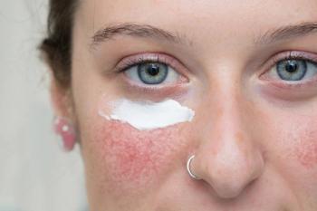
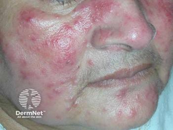
The evaluation was performed in six patients with clinical suspicion of having a Demodex-related disease, including three individuals with pityriasis folliculorum and three with papulopustular rosacea. All of the patients had undergone previous SSSB that documented the presence of >5 mites/cm2.
As reported by Dr. Fernandez-Nieto and colleagues, under FAV the Demodex mites were identified within the hair infundibulum as “roundish bright structures” when they were vertically oriented. Mites positioned at an oblique angle were seen as tubular dark structures surrounded by a whitish halo. The researchers note that the latter description corresponds to the appearance of the mites on RCM and that the whitish halo represented the mite’s exoskeleton.
Adjustment of the FAV system’s penetration depth made it possible to visualize the mite’s opisthosoma and gnathosoma, including the gut cells within the opisthosoma, and to estimate mite length.
“The FAV system allows counting of individual mites. With its field of view of 340 micrometers, mite density can be estimated quickly with just a few movements of the probe,” Dr. Fernandez-Nieto says. “We believe it is an equally reliable but easier and faster method than RCM or SSSB to evaluate mite density.”
Disclosure: Dr. Fernandez-Nieto has no relevant financial interests to disclose.
Reference:
1. Fernandez-Nieto D, et al. J Eur Acad Dermatol Venereol. 2020. Sep 14. Epub ahead of print.
Articles in this issue
over 5 years ago
Mohs micrographic presents challenges in kidsover 5 years ago
Adjunctive examined for facial erythemaover 5 years ago
Caffeine's role in skincareover 5 years ago
Strategies for choosing and using neuromodulatorsover 5 years ago
Robotic-controlled laser advances body contouringover 5 years ago
Gene expression profile test predicts metastatic riskover 5 years ago
Smartphone apps not effective in skin cancer riskover 5 years ago
New vehicles improve tolerability, acceptabilityover 5 years ago
HIPAA, COVID-19 and jailover 5 years ago
Treatment improves QOL for patients with acneAdvertisement
Related Content
Advertisement


Latest CME


Advertisement
Advertisement
Trending on Dermatology Times
1
New Review Confirms How Nonmedicated Skin Care Vehicles Drive Clinical Improvement
2
Zasocitinib Shows High Skin Clearance at Hard-to-Treat Psoriasis Sites
3
Dermatology Times Spot Test: July 19, 2026
4
Social Media Mythbusters: The Summer Skin Care "Shutdown"
5