

|Articles|November 12, 2018
- Dermatology Times, November 2018 (Vol. 39, No. 11)
- Volume 39
- Issue 11
Leg ulcers that require punch biopsies
Author(s)Dermatology Times Staff
Victor Desmond Mandel, M.D. and colleagues describe a reconstruction strategy for chronic infected neoplastic ulcers of the lower extremities using a dermal matrix and a unique postoperative dressing protocol.
Advertisement
“We believe that clinicians should be aware of the importance of an early and correct diagnosis of cutaneous neoplasms underlying chronic leg ulcers. This could avoid diagnostic delay and guarantee the best therapeutic approach to each patient,” they wrote.
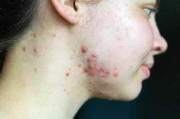
Lesions that grow, spread or are pigmented may require a biopsy and histologic study. If the wound doesn’t heal within three months of treatment, that may indicate the presence of a neoplasm requiring a skin biopsy and histologic study.
In the Sept. 11 issue of the Journal of Wound Care (
In one of two cases featured in the case report, they describe a 46-year-old male patient with laminin-332-deficient non-Herlitz junctional epidermolysis bullosa (JEB-nH).
Four months prior, the patient was admitted to the hospital with a 10x8 cm non-healing exophytic lesion on the right malleolus with visible scleroatrophic areas, erosions and hyperkeratosis, but no signs of acute infection.
A biopsy confirmed squamous cell carcinoma. A surgical excision was conducted on the exophytic lesion followed by reconstruction with a dermal matrix and silicone.
After three days, the matrix became infected (image A) and was removed. The surgeons then performed a single surgical hydrodebridement of the wound bed and a new graft with a dermal matrix (image B), followed by a post-operative treatment regime consisted of several layers of dressings in order to prevent surgical site infection. In this case a skin graft was impossible to perform because of the genetic condition. Image C shows the results achieved one month after reconstruction with dermal matrix and the post operative dressing.
“In our patients, the use of dermal matrix in conjunction with our dressing protocol resulted in complete wound healing. We achieved perfect engraftment of the matrix, almost complete restoration of the missing volume and good quality of grafted skin,” Dr. Mandel and colleagues wrote.
Articles in this issue
over 7 years ago
Low-dose bleomycin injections result in curious side effectover 7 years ago
7 Safety recommendations for tattoo removalover 7 years ago
Being older helps skin heal with less scarringover 7 years ago
A guide to wound careover 7 years ago
Topical sirolimus shows positive results in two studiesover 7 years ago
Wound healing in psoriasis, multiple sclerosisover 7 years ago
Classifying diabetic foot ulcersover 7 years ago
Role of protease targets in wound healing uncertainover 7 years ago
Allograft Tissue Research Grant program taking applicantsover 7 years ago
Verrica develops a solution for common wartsAdvertisement
Related Content
Advertisement


Latest CME


Advertisement
Advertisement
Trending on Dermatology Times
1
FDA Accepts Addition of Bemotrizinol as First New Sunscreen Ingredient in 20 Years
2
AI Analysis Reveals Menopausal Hair Loss Linked to Systemic Health and Inflammation
3
Dual-Action Microneedle Patch Shows Promise for Psoriasis in Preclinical Study
4
Novel Botanical Moisturizer Demonstrates Superior Efficacy Over Metronidazole in Phase 2 Rosacea Trial
5