

Publication|Articles|May 17, 2024
- Dermatology Times, May 2024 (Vol. 45. No. 05)
- Volume 45
- Issue 05
Dermatophytoma: A Diagnosis That You Do Not Want to Miss
Author(s)Shari R. Lipner MD, PhD
It is important dermatologists learn to recognize the clinical findings of dermatophytomas and distinguish them from other onychomycosis subtypes because the recommended treatments differ.
Advertisement
What Is a Dermatophytoma?
Dermatophytoma is a common yet highly underrecognized onychomycosis subtype that was first described by Roberts and Evans in the British Journal of Dermatology in 1998. Roberts and Evans described the clinical findings as a “dense white linear area or a round white area,” and when they clipped back the onycholytic area, they observed a “thick hyperkeratotic mass” that was not adherent to the nail plate and could be easily removed. The authors described the histopathology of the clipping as a “densely packed clump of dermatophyte hyphae, which are thick walled and somewhat abnormal looking.” They coined the term dermatophytoma, and likened it to an aspergilloma, which is also comprised of densely packed fungal elements. Roberts and Evans hypothesized that a nail unit dermatophytoma would have the same treatment implications as treatment of a lung aspergilloma, and that antifungal penetration and drug concentrations would be inadequate for a complete cure. Roberts and Evans recommended surgical removal of the nail plate, akin to surgical excision of a lung aspergilloma.1
Why Is It So Important to Recognize Dermatophytomas?
More than 25 years later, there have only been 40 papers that mention dermatophytoma as a key word, the majority of which are irrelevant or review papers. Therefore, although it is commonly seen in clinical practice, clinical findings and treatment are not well described. However, the reason that it is important that dermatologists learn to recognize the clinical findings of dermatophytomas and distinguish them from other onychomycosis subtypes is that the recommended treatments differ and affect patient outcomes.
What Are the Key Diagnostic Criteria for Dermatophytoma?
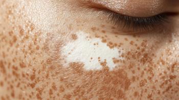
Dermatophytoma clinically presents as a yellow or white streak or patch involving the nail plate and typically affects the great toenail, although any nail may be affected (Figure). Dermoscopy, particularly contact dermoscopy using ultrasound gel, may be helpful to define the color and morphology, especially in equivocal cases. However, clinical examination alone is not sufficient to make the diagnosis of dermatophytoma. A microscopic examination with potassium hydroxide staining is needed to see the fungal hyphae packed into a fungal ball, or a histopathology on nail clippings would show densely packed fungal in mycelial configurations.
What Is the Recommended Treatment for Dermatophytoma?
Data on treatment of dermatophytomas are extremely limited, with studies being case series and retrospective studies. There have only been 4 prospective studies, all of which are open-label studies without a comparator. Sample sizes in dermatophytoma studies are small, end points vary between studies, and treatment courses are variable. Furthermore, recurrence rates are rarely reported. The reason that there are so few studies on dermatophytoma treatment is likely because most clinical trials exclude trial participants with dermatophytomas, potentially because they are historically challenging to treat, and/or because of the failure to recognize the distinct clinical presentation.
The most reported treatments for dermatophytoma are oral terbinafine and topical efinaconazole 10%. Topical luliconazole 5% and systemic itraconazole, fluconazole, and fosravuconazole have also been used. Surgical and chemical nail excision/debridement have been used alone or in combination with a topical or systemic antifungal.
Recommended treatment for dermatophytoma has evolved since it was first described in 1998. Early on, surgical excision of the dermatophytoma was recommended, with or without an oral or topical antifungal. However, in the past few years, several studies have demonstrated excellent efficacy with topicals alone. Because cure rates with combination treatment of surgical excision and oral terbinafine are higher than with oral terbinafine monotherapy, treatment with oral terbinafine alone is not recommended for dermatophytomas. To date, the largest studies on the treatment of dermatophytomas are with topical efinaconazole and topical tavaborole.
Of 223 participants with onychomycosis treated with efinaconazole 10% solution who were enrolled in a previous study, 82 participants with longitudinal spikes were evaluated in a post hoc analysis. The study authors found that the opacity ratio of longitudinal spikes decreased from 8.1 to 0.9 with treatment, indicating that there was resolution of the spikes. In addition, the complete cure rate for the whole nail, not just the spikes, was 41.5%, and the mycological cure rate was 69.5% following treatment. It should be noted that the onychomycosis cases were confirmed mycologically in the original study, but the longitudinal spikes were not confirmed histologically as densely packed hyphae.2
Of 366 participants with onychomycosis from a phase 2 study treated with topical tavaborole solution, 102 cases of dermatophytoma were analyzed in a post hoc analysis. Overall, 21 of 86 (24.4%) participants treated with tavaborole achieved complete resolution of dermatophytomas by day 180, compared with 0 of 16 (0%) participants treated with a vehicle. However, in this study, similar to the efinaconazole study, onychomycosis cases were confirmed mycologically in the original study, but the dermatophytomas spikes were not confirmed histologically as densely packed hyphae.3
Conclusions
A dermatophytoma is an important onychomycosis variant. Key diagnostic features are white or yellow streaks or patches in the nail plate, with hyphae aggregating in mycelial fashion of the histopathology of nail plate clippings. The recommended treatment has completely changed since it was first described more than 25 years ago, with topical therapy being the treatment of choice now. Keep in mind that a dermatophytoma may present concurrently with distal lateral subungual onychomycosis involving multiple nails, and such patient’s treatment with a systemic antifungal is indicated with topical treatment targeted at the dermatophytoma.
Shari R. Lipner, MD, PhD, is an associate professor of clinical dermatology, associate attending physician, and director of the Nail Center at the New York-Presbyterian Hospital/Weill Cornell Medical Center in New York, New York. She is a member of the American Academy of Dermatology, Women’s Dermatologic Society, Council for Nail Disorders, and American Dermatological Association, and is chair of multiple committees. She is the former president of the Dermatologic Society of Greater New York and is the president of the New York State Society of Dermatology and Dermatologic Surgery.
Financial disclosures: Dr Lipner has served as a consultant for Ortho Dermatologics, BelleTorus Corporation, Eli Lilly and Company, and Moberg Pharma.
Dr Lipner has no conflicts of interest relevant to the content of this submission.
References
1. Roberts DT, Evans EG. Subungual dermatophytoma complicating dermatophyte onychomycosis. Br J Dermatol. 1998;138(1):189-190. doi: 10.1046/j.1365-2133.1998.02050.x
2. Watanabe S, Iozumi K, Abe M, et al. Clinical effectiveness of efinaconazole 10% solution for treatment of onychomycosis with longitudinal spikes. J Dermatol. 2021;48(10):1474-1481. doi:10.1111/1346-8138.16035
3. Aly R, Winter T, Hall S, Vlahovic T. Topical tavaborole in the treatment of onychomycosis complicated by dermatophytoma: a post-hoc assessment of phase II subjects. J Drugs Dermatol. 2018;17(3):347-354. https://pubmed.ncbi.nlm.nih.gov/29537453/
Articles in this issue
about 2 years ago
Discovering Dermatology Times: May 2024about 2 years ago
Pearls From the Diversity in Dermatology Conferenceabout 2 years ago
Revealing Ruxolitinib’s Potential in Pediatric AD Treatmentabout 2 years ago
Unlocking Optimal AD Treatment: From Triggers to JAK Inhibitorsabout 2 years ago
Prebiotic Skin Care’s Effectiveness in AD for SOC PatientsAdvertisement
Related Content
Advertisement


Latest CME


Advertisement
Advertisement
Trending on Dermatology Times
1
Bempikibart Shows Positive Phase 2a Results in Alopecia Areata
2
Study Suggests Non-Ablative Fractional Laser Reverses Epigenetic Signatures of Skin Aging
3
The Gut-Skin Axis: New Evidence Links Microbiota to Skin Disease via Inflammatory Cytokines
4
Roundtable Discussion Weighs Access and Adherence Challenges in Vitiligo Care
5