

|Articles|November 8, 2019
- Dermatology Times, November 2019 (Vol. 40, No. 11)
- Volume 40
- Issue 11
Considerations for psoriasis patients with skin of color
Author(s)Ingrid Torjesen
Listen
0:00 / 0:00
Key Takeaways
- Psoriasis in skin of color can lead to pigmentary changes, affecting quality of life and requiring careful management and patient counseling.
- Erythema in darker skin tones may appear as violaceous, grey, or red-brown, complicating diagnosis and increasing misdiagnosis risk.
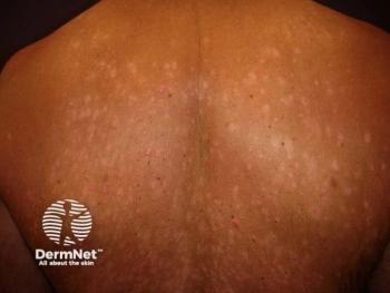
Psoriasis often has a greater impact on quality of life in patients with skin of color as the condition and its treatment can result in long-lasting pigmentary changes.
Advertisement
Psoriasis often has a greater impact on quality of life in patients with skin of color as the condition and its treatment can result in long-lasting pigmentary changes. Dermatologists should keep this in mind when managing the disease in this population to ultimately ensure that patients are fully aware of the risk of pigmentary changes and how they can be minimized, says Andrew Alexis, M.D., M.P.H., chairman of the department of dermatology and director of the Skin of Color Center at Mount Sinai St. Luke’s and Mount Sinai West in New York.
When assessing patients with skin of color for psoriasis, physicians should also be aware that clinical features can differ depending on the individual’s skin pigmentation, warns Dr. Alexis. In particular, erythema will be more difficult to perceive on darker Fitzpatrick V and VI skin tones.
TIPS FOR DIAGNOSIS
“The eye needs to be calibrated, so to speak, for the skin type of the patient because the redness of the lesions will be potentially masked by background melanin pigment in the epidermis,” he explains. So rather than a red or pink hue, erythema may appear violaceous, grey or red brown, depending on the background skin color.
Failing to appreciate the different appearance of erythema in skin of color raises the risk of confusing psoriasis with lichen planus, sarcoidosis or discoid lupus, especially the hypertrophic variant.
As well as the risk of misdiagnosis, he adds, “under-appreciation or under detection of the erythema component can lead to incorrect assumptions about the severity of the lesions.” This is important not only for the management of the condition in a patient but also for clinical trials where erythema is formally measured as an EASI score or SCORAD to assess treatment efficacy.
So, what are his tips for diagnosis?
- Expand your color palette to detect different ranges of erythema. Think about how psoriasis typically looks across different skin types, include violaceous hue, red brown hue and greyish hue. Then, rely on other clues such as the quality of the scale to confirm the diagnosis.
- Look at how well the lesion is demarcated. In psoriasis, lesions are usually sharply demarcated.
- Look at anatomic distribution of the lesions. This can help point to the correct diagnosis, evenwhen the erythema may be difficult to perceive.
RISK OF PIGMENTARY CHANGES
Inflammatory disorders of the skin such as psoriasis can be associated with post inflammatory pigment alterations, which raises an important consideration when treating patients with skin of color.
“Not only does one have to counsel the patient on management of psoriasis plaques themselves, but also the pigmentary changes that may result from the psoriasis plaques,” says Dr. Alexis. “A plaque may resolve with hyperpigmentation or hypopigmentation that can be just as impactful for the patient as the psoriasis lesion itself. This needs to be included in the discussion with the patient.
“If the psoriasis starts to resolve and leaves behind a dark or light-colored spot, the patient may either feel that the treatment is not working or making the condition worse.”
Pigmentary alterations may resolve with time, but some patients will find them particularly bothersome and want them dealt with more quickly. Once the psoriasis has resolved, hyperpigmentation can be treated with a topical retinoid to manage both psoriasis and hyperpigmentation or with a formula containing topical hydroquinone, such as modified Kligman Willis, which contains hydroquinone, retinoid and a corticosteroid.
For patches of hypopigmentation, lightbased therapies are an option for speeding up the return of pigments.
“I believe that, if we effectively manage the underlying psoriasis, we may be able to reduce the severity and duration of pigmentary alteration,” says Dr. Alexis. “So, it makes it especially important not to undertreat this disease in darker skinned patients where there is the additional burden of pigmentary alterations.”
TREATMENT CONSIDERATIONS
The treatment approach for this group is similar to treatment for lighter skinned patients, but with a few nuances.
For example, while hypopigmentation secondary to corticosteroid is a risk for all patients, its impact is greater and more debilitating in darker skinned patients. The duration of corticosteroid use should, therefore, be limited, and clinicians should ensure that patients fully understand where and how to apply them and not to over apply in order to minimize the risk of hypopigmentation, Dr. Alexis says.
When it comes to choice of other topical therapies, tolerability is important because irritant dermatitis secondary to the topical therapy could induce more hyper- or hypopigmentation, he adds.
Use of phototherapy and the excimer laser risks worsening hyperpigmentation, so patients need to be warned about this, says Dr. Alexis, and also that phototherapy will promote diffuse tanning of the skin. Patients may not expect this overall darker skin tone, and it may not match their personal or cultural view of what their skin color should be, he explains.
There are specific considerations for choosing a topical regimen for the treatment of scalp psoriasis, particularly for women of sub-Saharan African ancestry with Afro-textured hair, because of the structural differences of the hair and the haircare practices they may follow. It is important to get the patient’s input, says Dr. Alexis. “One has to take into account their haircare practices and come up with the regimen that is compatible. Hair washing frequency may differ - it tends to be less frequent - and the vehicle has to be compatible with their overall hairstyle and hair structure.”
For example, if daily hair washing with a medicated shampoo is recommended for someone with Afro-textured hair that has been straightened with a chemical relaxer, frequenthair washing may lead to dryness, fragility of hair, breakage or it simply may not be practical given the time-consuming after-shampoo haircare routine.
These considerations will impact choice of vehicle and dosing frequency of a topical and even the threshold for using a non-topical therapy, he says.
“I have a lower threshold for using systemic therapies because of the impact of scalp psoriasis on this cohort,” he admits, and says there is also an argument for having a lower threshold for initiating systemic therapies for plaque psoriasis.
“Given the high rate of pigmentary alterations with topicals, which can be long-lasting and disfiguring, appropriate use of systemic agents is key,” he says. “In other words, avoiding undertreatment. With undertreatment would come a greater tendency for long-standing pigmentary alterations on top of the long-standing issues of psoriasis itself.”
References:
1. Xiao Y, Zhang X, Luo D, Kuang Y, Zhu W, Chen X, Shen M. The efficacy of psychological interventions on psoriasis treatment: a systematic review and meta-analysis of randomized controlled trials. Psychology Research and Behavior Management. 2019; 12: 97–106; doi: 10.2147/PRBM.S195181.
2. Ferreira BI, Abreu JL, Reis JP, Figueiredo AM. Psoriasis and Associated Psychiatric Disorders: A Systematic Review on Etiopathogenesis and Clinical Correlation. J Clin Aesthet Dermatol. 2016 Jun; 9(6): 36–43.
Articles in this issue
over 6 years ago
Phototherapy safe, effective for psoriasisover 6 years ago
Technology improves histologic diagnosis in melanomaover 6 years ago
AI: It’s not dermatologist vs. machineover 6 years ago
What's new in skincareover 6 years ago
Cosmetic technology advancesover 6 years ago
Melanoma management perspectives varyover 6 years ago
Fat may play a protective role in acneover 6 years ago
Global panel updates rosacea recommendationsover 6 years ago
Do patients have property rights in healthcare?over 6 years ago
Novel acne cream seeks FDA approvalAdvertisement
Related Content
Advertisement


Latest CME


Advertisement
Advertisement
Trending on Dermatology Times
1
Real-World Study Finds Experienced Dermatologists Outperform AI in Skin Cancer Diagnosis
2
New Review Confirms How Nonmedicated Skin Care Vehicles Drive Clinical Improvement
3
Dermatology Times Spot Test: July 19, 2026
4
Social Media Mythbusters: The Summer Skin Care "Shutdown"
5