

|Articles|January 1, 2005
Avoid trauma in PG care
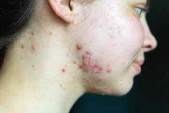
Part of the problem is that PG is very uncommon and can be difficult to diagnose.
Advertisement
"PG can present a diagnostic and therapeutic challenge and so can be a frustrating problem for both the patient and physician," she says. "Yet those same features can make it a very rewarding entity to treat because there is the opportunity to achieve healing in a long-suffering patient," says Dr. Parish, clinical instructor, department of dermatology and cutaneous biology, Jefferson Medical College, Thomas Jefferson University, Philadelphia.
"With sufficient time, resolution can often be achieved using a variety of topical, intralesional, and systemic therapies, but please, no surgery!" she cautions.
"Recently, one patient came to me for another opinion after bilateral leg amputation had been recommended," she tells Dermatology Times.
"That individual had ulcers measuring 20 x10.5 cm on each leg, and unfortunately was not the first pat-ient I had seen whose condition was worsened because of misdiagnosis or misunderstanding of the care of this ulcer by another physician," Dr. Parish says.
Difficult diagnosis Part of the problem is that PG is very uncommon and can be difficult to diagnose. Extreme pain and pres-ence of a violaceous, undermined border are important clinical clues.However, PG identification requires exclusion of other entities of similar appearance, including vascular disorders, an infectious etiology, drug-induced ulcers, neoplasms, and factitial ulcers. Therefore, the work-up should include wound culture, history and physical exam. In addition, a variety of laboratory, imaging, and other special studies may be undertaken to diagnose an associated systemic disease.
"The identification of PG in part requires a search for an underlying disease process, which is present in 50 percent of patients. Inflammatory bowel disease, polyarthritis, and hematologic diseases are most common, but only a very small percent of patients with those problems develop PG, and PG has rarely been associated with a long list of other systemic diseases," Dr. Parish says.
A role for lesion biopsy in the diagnostic evaluation is questionable considering the risk of a pathergic phenomenon. The histopathology of PG is nonspecific - early lesions are characterized by a primarily neutrophilic infiltrate involving the follicular structures, and more developed lesions show ulceration, necrosis and a mixed cell infiltrate.
Nevertheless, biopsy is useful because it helps rule out other entities, and Dr. Parish believes it should be undertaken to help establish the diagnosis of PG considering that most of the treatments for PG are associated with significant safety concerns.
Therapeutic decisions Once the diagnosis is made, therapeutic decisions may be influenced by lesion size and any comorbid disease.
Choices for topical and intralesional therapy can include corticosteroids, tacrolimus, disodium chromoglycate, nicotine and cyclosporine. By itself, however, local treatment usually is not adequate and systemic therapy is required.
Historically, steroids and various other immunomodulating agents have been used. Steroid treatment requires a high dose (eg., prednisone 1-2 mg/kg/day) and often prolonged treatment. Therefore, Dr. Parish notes that she now prefers to start immunosuppressive therapy with cyclosporine A 3-5 mg/kg/day in patients who have no medical contraindications to its use. However, there have been a number of recent reports describing success with the TNF-alpha inhibitor infliximab (Remicade, Centocor), and that may be a particularly rational choice in patients with comorbid rheumatoid arthritis or inflammatory bowel disease.
Advertisement
Related Content
Advertisement


Latest CME


Advertisement
Advertisement
Trending on Dermatology Times
1
FDA Accepts Addition of Bemotrizinol as First New Sunscreen Ingredient in 20 Years
2
Dual-Action Microneedle Patch Shows Promise for Psoriasis in Preclinical Study
3
AI Analysis Reveals Menopausal Hair Loss Linked to Systemic Health and Inflammation
4
Health Canada Grants Priority Review to Birch Triterpenes for EB
5