

|Articles|December 13, 2019
- Dermatology Times, February 2020 (Vol. 41, No. 2)
- Volume 41
- Issue 2
Tape strips may replace biopsies in pediatric atopic dermatitis
Author(s)Ingrid Torjesen
Biopsies are the gold standard approach for obtaining skin samples to test for biomarkers in atopic dermatitis, but tape strips may offer a far less invasive alternative for young children with the disease, research suggests.
Advertisement
Biopsies are the gold standard approach for obtaining skin samples to test for biomarkers in
Skin profiling studies have associated certain biomarkers with the development of atopic dermatitis, and these biomarkers can help physicians assess disease severity and determine the best treatment approach for each patient.2,3 However, performing a skin biopsy is often not practical in young children. While blood analysis is also less invasive, it cannot capture the whole atopic skin phenotype, which has led researchers to search for other techniques for younger patients, including tape strips. To date, most studies have focused on testing such strips in adults with atopic dermatitis.
For this study, published in October in JAMA Dermatology, researchers from several institutions performed mRNA profiling on the tape strips used to gather samples from children with moderate-to-severe atopic dermatitis of less than six months duration.
The strips were used on a total of 51 children younger than five years old at the Ann & Robert H. Lurie Children’s Hospital of Chicago. Strips were applied to the skin of 21 lesional and non-lesional children who had developed atopic dermatitis within the last six months, and 30 children who did not have atopic dermatitis. Gene and protein expression were then evaluated using real-time polymerase chain reaction and immunohistochemistory.
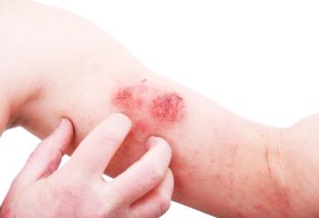
Each child had 16 large D-Squame tape strips (CuDerm Corp) applied. Lesional skin was collected from the antecubital fossa (i.e., the triangular region in the forearm on the anterior surface of the elbow) when possible, and non-lesional skin was sampled from nearby on the same arm. Skin from children without atopic dermatitis was sampled from the same areas.
One of the study authors, Emma Guttman-Yassky, M.D., Ph.D., at the Icahn School of Medicine at Mount Sinai explains it was “crucial to understand whether profiling of the tiny amounts of RNA obtained from the outer layers of the skin using tape strips can accurately yield key biomarkers for AD."
Against a panel of 79 genes, 77 immune and barrier gene products were detected (gene detection rate 97%) from 70 of 71 tape strip samples (sample detection rate, 99%). Significant differences were seen in detection of 53 of the markers between children with lesional and/or nonlesional atopic dermatitis and children without the skin condition.
Many cellular markers of T-cells (CD3), atopic dermatitis-related dendritic cells (Fc ε RI and OX40 ligand receptors), and key inflammatory (matrix metallopeptidase 12), innate (interleukin 8 [IL-8] and IL-6), helper T-cell 2 (TH2; IL-4, IL-13, and chemokines CCL17 and CCL26), and TH17/TH22 (IL-19, IL-36G, and S100A proteins) genes were significantly increased in samples taken from atopic dermatitis patients compared with those from normal skin. Conversely there were decreases in other markers including epidermal barrier gene products (FLG, CLDN23, and FA2H) and negative immune regulators (IL-34 and IL-37).
Several of the biomarkers had not been detected or evaluated in previous atopic dermatitis tape strip studies, including measures of cellular infiltrates such as those associated with T-cells and T-cell activation (CD3 and ICOS), atopic dendritic cells (Fc ε RI and OX40L), and key inflammatory markers of general inflammation (MMP12) and innate immunity (IL-8 and IL-1RA), and epidermal proliferation (SERPINB3).
Dr. Guttman-Yassky tells Dermatology Times, “We are constantly looking to develop non-invasive biomarkers to track skin improvement in children, but also in adults.”
As the use of tape strips is painless and nonscarring, it would be useful for longitudinal pediatric studies and clinical trials, in which serial measures are needed to identify predictors of response, course, and comorbidities, the researchers conclude.
“I do believe that in the future such tests may be commercially available. Right now, they are done by specialized labs like mine, as they require very skilled personnel, but I can see more general applications in the future,” Dr. Guttman-Yassky adds.
References:
1. Guttman-yassky E, Diaz A, Pavel AB, et al. Use of Tape Strips to Detect Immune and Barrier Abnormalities in the Skin of Children With Early-Onset Atopic Dermatitis. JAMA Dermatol. 2019;
2. Brunner PM, Israel A, Zhang N, et al. Early-onset pediatric atopic dermatitis is characterized by T2/T17/T22-centered inflammation and lipid alterations. J Allergy Clin Immunol. 2018;141(6):2094-2106.
3. Esaki H, Brunner PM, Renert-yuval Y, et al. Early-onset pediatric atopic dermatitis is T2 but also T17 polarized in skin. J Allergy Clin Immunol. 2016;138(6):1639-1651.
Articles in this issue
over 6 years ago
Gene expression profile testing aids skin cancer managementover 6 years ago
How to diagnose rosacea in skin of color patientsover 6 years ago
Rosacea commonly underdiagnosedover 6 years ago
Technology targets non-genital wartsover 6 years ago
Can sweat shed a light on disease severity in atopic dermatitis?over 6 years ago
Adjunctive platelet-rich plasma may improve acne scarsover 6 years ago
An investigational drug for keloidsover 6 years ago
Study supports evidence on indoor tanning riskover 6 years ago
Nano-pulse stimulation promising for skin lesionsover 6 years ago
Mind-body therapies effective for skin disordersAdvertisement
Related Content
Advertisement


Latest CME


Advertisement
Advertisement
Trending on Dermatology Times
1
Real-World Study Finds Experienced Dermatologists Outperform AI in Skin Cancer Diagnosis
2
New Consensus Defines Remission, Disease Activity in Atopic Dermatitis
3
Beyond Retinol: Evaluating the Stability, Safety, and Gene-Expression Profile of Bakuchiol Ferulate
4
Zasocitinib Shows High Skin Clearance at Hard-to-Treat Psoriasis Sites
5