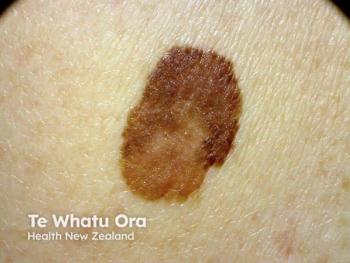
Total immersion photography may catch more melanomas at noninvasive stage
Total immersion photography, performed with the Melanoscan device (Melanoscan Inc.), has the potential to reduce operator error and possibly catch nearly all melanomas in the noninvasive stage, says an expert.
Las Vegas - Total immersion photography, performed with the Melanoscan device (Melanoscan Inc.), has the potential to reduce operator error and possibly catch nearly all melanomas in the noninvasive stage, says an expert.
“The real problem with total body photography (TBP) is that it depends on the photographer,” says Rhett Drugge, M.D., a dermatologist and cosmetic surgeon based in Stamford, Conn., and developer of the Melanoscan. Dr. Drugge spoke at the Cosmetic Surgery Forum in December.
Traditional TBP aims to create a standard array of clinical images shot from various perspectives, he explains. With the photographer shooting images of a mostly stationary patient, he adds, shadowing and camera angle discrepancies create inconsistencies. For such reasons, the United States Preventive Services Task Force says, standard TBP has not shown an adequate risk-benefit ratio to support its use in melanoma screening (Wolff T, Tai E, Miller T. Ann Intern Med. 2009;150(3):194-198.).
Likewise, conventional skin exams performed with the naked eye offer simplicity but may miss early melanomas, Dr. Drugge says. For example, he says physicians may be unable to perceive atypia in a lesion measuring less than 3 mm in diameter. However, “That’s the size at which most people would like to find melanomas; we’ve discovered that those are primarily melanoma in situ (MIS), for which the cure rate approaches 100 percent.”
To that end, “The data appear to demonstrate that, using the Melanoscan as often as annually, we can drive up the rate of early melanoma detection by highlighting growth or change characteristics of people’s lesions over time.”
Device specs
The device features mounted cameras (added to a standard phototherapy booth) that shoot all the required images without having to rearrange the equipment, he says.
“As digital imaging has become more cost-effective, having an imaging array becomes an attractive and highly reproducible method for achieving TBP with highly standardized perspectives, lighting and other parameters,” he says.
The 25-camera array fires simultaneously, “And we reposition people to optimize the display of all their underarm skin,” plus skin folds and other areas where melanomas may hide. The patient spends around 2.5 minutes in the booth, says Dr. Drugge, who admits that the procedure is equipment-intensive. The device costs around $62,000, including the phototherapy booth.
However, a well-trained nurse or aesthetician can perform the scan and prepare a report highlighting any changes observed in a patient’s lesions. “Then I review the reporting, fully examine the patient and come up with my results,” which could be that a suspicious-looking lesion wasn’t actually melanoma, or vice versa.
With the Melanoscan, “We can process five to 10 people an hour, capture and store all that imagery and make it available for mapping of biopsy specimens, dermoscopy elements, pathology requests and prior images of the patient’s entire body.”
Key comparisons
A key study compared the performance of a Melanoscan-equipped clinic against that of three clinics without the device from 2005 to 2009.
“It got us interested in whether the MIS ratio is somehow being changed in our setting, because we observed an enrichment of our MIS ratio,” Dr. Drugge says. Specifically, the MIS/invasive melanoma ratio at the non-Melanoscan practices fell between 1.0 and 1.3, versus 1.625 in the Melanoscan clinic (Stricklin SM, Stoecker WV, Malters JM, et al. J Am Acad Dermatol. 2012;67(3):e105-109.).
Based on this observation, Dr. Drugge says, “I hypothesized that the scanning intervention - and the frequency of scanning - were driving the ratio.” In this regard, “We broke down the data to show that there’s about an 82.5 percent MIS ratio when people were scanned annually, versus 85 percent when scanned within 11 months (Rhett Drugge, M.D., Presented at Internet Dermatology Society/IDS Annual Meeting. Sept. 21, 2012. Austin, Texas).”
Moreover, he says, about 40 percent of the melanomas diagnosed by the average U.S. physician are MIS. The ratio at the three comparator practices that did not use Melanoscan was approximately 50 percent, he says.
Conversely, “In our group, we were driving more toward 90 percent at a six-month scan interval, and the ratio will probably be rising.” As scanner sensitivity and frequency increase going forward, he says, “I hypothesize that if we are really good with screening, all melanomas can be caught in the epidermis, and therefore virtually 100 percent curable.”
Next steps
Accordingly, “The next phase for the Melanoscan is to try to make the resolution even greater, so we can be more sensitive to the very earliest melanomas. In fact, we have a statistically insignificant but growing population of melanomas followed up every six months, in which we are showing a trend toward 88.5 percent of melanomas being MIS (not yet submitted for publication).”
Some dermatologists believe that invasive melanomas originate in the dermis rather than the epidermis. In contrast, Dr. Drugge says, “Our data seem to suggest that they come from the epidermis, then invade the dermis in a predictable progression” over a period of three to five years.
By the same token, Dr. Drugge and colleagues have shown that the average Breslow thickness of melanomas in patients followed with Melanoscan increased from less than 0.2 mm at an interval of one or two years, to 3 mm at a three-year interval (Elizabeth Drugge, Ph.D., M.P.H. Presented at IDS Annual Meeting. Feb. 3, 2011. New Orleans).
The same study also showed that the vast majority of invasive tumors found in 68 patients who underwent Melanoscan serial screening, and in 82 patients traditionally screened, measured less than 1 mm deep. However, five traditionally screened patients had melanoma invasion up to 5 mm, versus none deeper than 1 mm in the Melanoscan group.
“The weakness of Melanoscan studies is that the scanned people are more compliant and more likely to undergo regular melanoma screenings,” as well as to conduct skin self-examinations, Dr Drugge says.
Next steps for the device include a multicenter trial to further validate the Melanoscan process, he says. Meanwhile, developers will continue refining its image-analysis algorithms.
Additionally, Dr. Drugge and his colleagues are considering adding approximately a dozen more high-value sites, such as academic centers and large organizations that can contribute to data gathering and the ongoing fine-tuning of the device. In this regard, however, “We don’t want to get too far ahead of ourselves” by pursuing new sites if doing so compromises the Melanoscan company’s ability to support its five existing sites. DT
Disclosures: Dr. Drugge is the developer and patent holder of the Melanoscan device.
Newsletter
Like what you’re reading? Subscribe to Dermatology Times for weekly updates on therapies, innovations, and real-world practice tips.


