- Dermatology Times, June 2020 (Vol. 41, No. 6)
- Volume 41
- Issue 6
Stem cell factor inhibition for hyperpigmented skin
Stem cell factor, a growth factor critical for melanocyte survival, may play an important role in the development of benign and malignant skin hyperpigmentation disorders.
Stem cell factor, a growth factor critical for melanocyte survival, plays an important role in the development of benign and malignant skin hyperpigmentation disorders. More research is needed to look at targeting stem cell factor inhibition as a way to treat skin lesions from melasma to melanoma, according to an article published December 2019 in the Asian-Pacific Journal of Cancer Prevention.
Stem cell factor binds to the receptor tyrosine kinase and is expressed by the body’s fibroblasts and endothelial cells, promoting melanocyte differentiation proliferation, migration and survival, according to the authors.
“The interaction of stem cell factor with its receptor, c-kit, is well known to be crucial for the survival of melanocytes,” the Egyptian and Saudi Arabian researchers write.
But little is known about the role of stem cell factor/c-kit interaction in epidermal pigmentation, according to the paper.
These researchers studied stem cell factor expression in benign and malignant pigmented lesions to determine the growth factor’s role in pathogenesis and potential use as a therapeutic target.
They studied stem cell factor expression in 60 patients with benign hyperpigmented lesions, including 20 with melasma, 20 with solar lentigines and 20 with freckles. They also studied 36 patients with hyperpigmented skin cancers, including 14 with basal cell carcinoma, 12 with squamous cell carcinoma and 10 who had malignant melanoma.
They found stem cell factor was positively expressed in all melasma, solar lentigines and freckled lesions.
More specifically, immunohistochemical testing revealed significantly higher stem cell factor expression in melasma lesions than in perilesional skin. They found moderate expression in 60% and strong expression in 40% of melasma cases in melasma lesions. While there was only weak expression in 100% of melasma cases in perilesional areas.
The intensity of stem cell factor expression in lentigines lesions was weak in 10%, moderate in 30% and strong in 60% of cases, while perilesional areas in those cases had no stem cell factor expression in 20% of patients and weak expression in 80%.
Stem cell factor expression intensity was increased in the lesional parts of freckles cases, with weak expression in 20% of patients, moderate in 30% and strong expression in 50%. Stem cell factor was not expressed in 20% of perilesional areas in patients with freckles, weakly expressed in 70% and moderately expressed in 10% of perilesional areas.
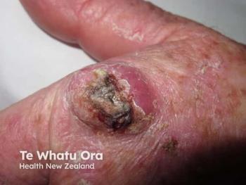
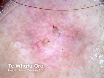
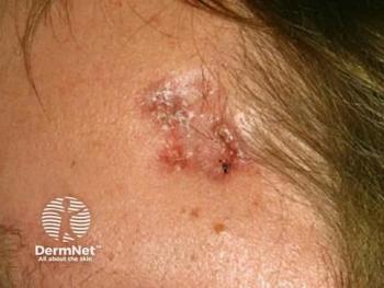
Stem cell factor expression was significantly higher in basal cell carcinoma tumor cells compared to normal surrounding keratinocytes. The researchers found the squamous cell carcinoma cases they studied showed 50% cytoplasmic positivity for stem cell factor expression, suggesting stem cell factor is implicated in squamous cell carcinoma pathogenesis. Stem cell factor was also positively expressed in malignant melanoma.
While basal cell carcinoma, squamous cell carcinoma and malignant melanoma showed variable intensities of cytoplasmic expression of stem cell factor, the increase in expression in basal cell carcinoma and melanoma tumor cells was statistically significant, the authors write.
The researchers concluded their findings strongly suggest stem cell factor plays a key role in the development of common benign and malignant skin hyperpigmented disorders, but more research is needed to clarify that role. Studies looking at using stem cell factor as a target for developing new treatments for hyperpigmented disorders and malignancies also are needed.
Disclosures:
The authors have no conflicts of interest, according to the paper.
References:
Atef A, El-Rashidy MA, Abdel Azeem A, Kabel AM. The Role of Stem Cell Factor in Hyperpigmented Skin Lesions. Asian Pac J Cancer Prev. 2019 Dec 1;20(12):3723-3728. doi: 10.31557/APJCP.2019.20.12.3723.
Articles in this issue
over 5 years ago
Pipeline progress in epidermolysis bullosaover 5 years ago
New treatments help improve rosacea managementover 5 years ago
Maintenance therapy for basal cell carcinoma proves effectiveover 5 years ago
How to perfect the cosmetic injectable consultationover 5 years ago
Laser therapy may be practical for some basal cell carcinomasover 5 years ago
Topical combination helps clear truncal acneover 5 years ago
Research gains traction for hidradenitis suppurativaover 5 years ago
Melanoma tumor syndromesover 5 years ago
New device treats nonmelanoma skin cancerNewsletter
Like what you’re reading? Subscribe to Dermatology Times for weekly updates on therapies, innovations, and real-world practice tips.


