
|Articles|November 1, 2017
A squamous cell carcinoma that disproportionately affects men
Author(s)Amy Reyes
Factors that drive lip squamous cell carcinomas in men.
Advertisement
Squamous cell carcinomas of the lip, tongue and floor of the mouth are each associated with unique expressions. Yet, they share one key factor: They disproportionately affect men. Researchers suggest this may be due to high tobacco use in this group as smoking is associated with 75 percent of these three squamous cell carcinomas.
When tobacco isn’t a factor, an individual’s genetic predisposition may play a role. People who have been treated for cancer with chemotherapy or radiation may be more susceptible to oral squamous cell carcinomas (oSCC) when DNA fails to repair itself after treatment.
Also, def ciencies in vitamins A, E or C may be factors as might defects in the immune system; the body’s inability to metabolize carcinogens; or, chronic sun damage. In this article, we narrow in on a case of lip squamous cell carcinoma in a 31-year-old man.
Lip SCC
Traditionally, the classification of oral squamous cell carcinoma (oSCC) has included lip squamous cell carcinoma (lSCC), but recent studies have shown that genetics and clinical behavior associated with lSCC is unique as compared to the oral and cutaneous forms of the condition.
Nour Kibbi, M.D.
For instance, while the human papillomavirus plays a role in oSCC, it has not yet been implicated in lSCC, which is known to be associated with sun exposure and the effects of immunosuppression, just as in cutaneous squamous cell carcinoma (cSCC).
Treatment is associated with high success rates. The estimated five-year survival rates following surgical treatment of lSCC is over 80 percent, compared with 65-70 percent for oSCC and 92 percent for cSCC.
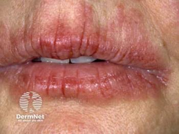
Lip squamous cell carcinoma can present as a verrucous or keratotic plaque, an exophytic papule or nodule, or as a non-healing ulcer. The lower lip is approximately 12 times more likely to be af ected due to greater exposure to sunlight.
“It is important for dermatologists to be aware of lSCC as a unique entity. Management options present unique challenges,” said Nour Kibbi, M.D., a resident physician in dermatology at the Yale-New Haven Medical Center in Connecticut.
Secondary malignancies
Patients who have had a hematopoietic stem cell transplant (HSCT) have a 2-6 percent higher risk of developing a secondary malignancy 10 years post treatment, and a 6-13 percent increase at 15 years. Adding male gender and a post-transplant chronic graft versus host disease (GvHD) to the mix heightens the risk, Dr. Kibbi said. Typically, leukemia or lymphoma is the most common secondary malignancy followed by SCC of oral cavity, skin or GI track. The risk for oSCC is 17 times greater than that of the average population.
Last month, during the American Society for Dermatologic Surgery annual meeting in Chicago, Dr. Kibbi presented a case study of 31-yearold man with lSCC who was successfully treated with Mohs micrographic surgery (MMS).
“SCC of lip can be a challenging diagnosis in the setting of complex medical history,” she said.
Case study: 31-year-old man
This was a 31-year-old man with a history of lymphoblastic lymphoma. He was treated with a HSCT, which was complicated by a chronic graft versus host disease. “Three years after the transplant, he developed a verrucous, sessile plaque on the right lower lateral vermilion lip of 2.5 x 1.7 cm extending from the vermilion border to the wet vermilion sparing the vestibule and other oral structures. He had no associated lymphadenopathy,” Dr. Kibbi stated in her presentation.
“The biopsy findings included atypical parakeratosis above acanthosis composed of atypical eratinocytes with a pushing border into the lamina propria; p16 immunohistochemistry was negative. The lesion was staged T2a by Brigham and Women’s criteria,” she stated.
Treatment options
Surgery and radiotherapy are standard treatment options for oral squamous cell carcinomas. There were several treatment options: Wide local excision (with 1-2 cm surgical margins), a lymph node dissection with or without radiation and MMS.
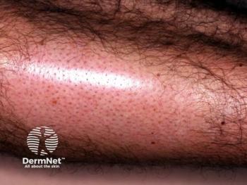
At the recommendation of Dr. Kibbi and her colleagues, Kathleen Suozzi, M.D., and David J. Lef ell, M.D., the patient opted for MMS. Three months after the surgery, he was left with a 5mm keratotic papule on the right lower vermilion. A biopsy revealed it was a hyperplastic actinic keratosis to base. It was treated with electrocautery to the base.
“This case adds to the sparse literature of five other cases of lip squamous cell carcinoma arising in the setting of GVHD. All six reported cases arose in relatively young males (average age 42). In particular, our patient, only 31 at the time of diagnosis, already had clinical actinic damage and solar elastosis, likely in part due to being on voriconazole prophylaxis, a known potentiator of UV-irradiation-induced damage. Second, all six cases had developed oral GVHD prior to lSCC; by upregulating cytokines (particularly type I interferons), GVHD is thought to enhance malignant transformation,” the authors wrote.
Mohs micrographic surgery
Mohs is associated with high cure rates. A prospective, multicenter Australian case series spanning 10 years reviewed 1,263 patients with squamous cell carcinoma who were treated with Mohs micrographic surgery. Researchers found a five-year recurrence rate of 3.9 percent. For recurring tumors, that rate jumped to 5.9 percent compared to 2.6 percent of primary tumor excisions. For patients with perineural invasion, the recurrence rates were 8 percent over five years.
REFERENCES
Rizzo JD, Curtis RE, Socie G, et al.
Bota JP, Lyons AB, Carroll BT. “Squamous Cell Carcinoma of the Lip-A Review of Squamous Cell Carcinogenesis of the Mucosal and Cutaneous Junction,” Dermatologic Surgery. April 2017. DOI:10.1097/DSS.0000000000001020.
Anastasios K Markopoulos.
“Squamous Cell Carcinoma of the Lip in a Patient with Graft-Versus-Host Disease,” Nour Kibbi, M.D., and Kathleen Suozzi, M.D. American Society for Dermatologic Surgery annual meeting, October 2017, Chicago.
Leibovitch I, Huilgol SC, Selva D, et al.
Vivek V. Gurudutt and Eric M. Genden. "Cutaneous Squamous Cell Carcinoma of the Head and Neck," Journal of Skin Cancer. Feb. 21, 2011. DOI:10.1155/2011/502723
Advertisement
Related Content
Advertisement

Latest CME


Advertisement
Advertisement
Trending on Dermatology Times
1
Social Media Mythbusters: Skin Cycling
2
Clinique's Daily Calm Merges Clinical Efficacy with Color Cosmetics for Sensitive Skin
3
First HA Injectable for Neck Wrinkles Cleared by FDA
4
What Dermatologists Need to Know About the First New Sunscreen Ingredient in Decades
5