

|Articles|August 1, 2018
- Dermatology Times, September 2018 (Vol. 39, No. 9)
- Volume 39
- Issue 9
Pros and cons of imaging technologies for melanoma
Author(s)Whitney J. Palmer
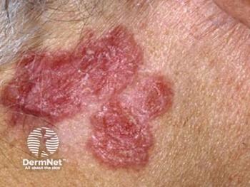
Monitoring changes in patients with melanoma can be tricky. Some changes present slowly and in tiny ways. Using imaging technologies to track these changes can help achieve an accurate diagnosis.
Advertisement
Monitoring changes in patients with melanoma can be tricky. Some changes present slowly and in tiny ways. Using imaging technologies to track these changes can help achieve an accurate diagnosis.
Jennifer Stein, M.D., Ph.D., associate director of the Pigmented Lesion Section in the Ronald O. Perelman Department of Dermatology at the New York University School of Medicine, discussed the use of digital imaging during the 2018 American Academy of Dermatology Summer Meeting.
Using imaging technologies can improve the specificity and sensitivity with which you treat patients, she says.
IMAGING OPTIONS
Total Body Photography (TBP): This method can help identify new changes in patients with atypical nevi for melanoma detection, she says. Being able to more accurately highlight problem spots can reduce the number of unnecessary biopsies. One study reported a 3.8-fold decrease in biopsies when TBP was used correctly, she says. In addition, TBP can help patients with self exams because they have previous visual images to make comparisons. Another study, she says, revealed patients were roughly 12 percent more likely to accurately identify changes when they had reference photos.
TBP can be recommended during 6-month check ups for all patients at high risk for melanoma. It best serves patients who have relatively easy nevi to manage.
Sequential Digital Dermoscopic Imaging (SDDI): SDDI is a more rigorous form of TBP generally completed on patients who have more aggressive forms of disease. It’s done in the short term, Stein says, lasting approximately 3 months. It requires the patient to return for more frequent visits.
SDDI is most appropriate for patients who have complex lesions.
PROS and CONS
There are pros and cons to TBP, she says. In addition to monitoring lesion changes, reducing biopsies, and improving self exams, TBP also decreases patient worry and helps patients who have difficulty tracking moles.
However, it is expensive, time consuming, can cause patient discomfort, and presents privacy issues if photos are stored in the general electronic health record.
ARTIFICIAL INTELLIGENCE
Convolutional neural networks are the next step in digitally monitoring melanoma progression in patients, Stein says. This technology can be trained, using several thousands of reference images, to recognize almost any condition.
While it won’t replace you as the dermatologist ultimately responsible for diagnosis and treatment, she says, it could potentially help you triage patients and identify high-risk lesions that need immediate attention. It could also be used to screen patients without easy access to a dermatologist, pinpointing which patients would actually need a dermatology referral.
REFERENCE
Jennifer Stein, M.D., Ph.D., “Advances in Imaging Technology for Melanoma Diagnosis.” American Academy of Dermatology 2018 Summer Meeting, Chicago, Ill., July 28, 10a.m.-1p.m.
Articles in this issue
almost 8 years ago
Spironolactone as effective as antibioticsalmost 8 years ago
Psychological stress as acne causealmost 8 years ago
Technology: A private practice’s best friendalmost 8 years ago
Lack of standardization?almost 8 years ago
Update: Immunotherapy for melanomaalmost 8 years ago
A new treatment for seborrheic keratosis examinedalmost 8 years ago
Hidradenitis suppurativa and Crohn’s disease linkalmost 8 years ago
Clinical trials recruiting: Molluscum contagiousumalmost 8 years ago
Psoriasis lotion seeks FDA approvalAdvertisement
Related Content
Advertisement


Latest CME


Advertisement
Advertisement
Trending on Dermatology Times
1
Stapokibart Successfully Treats Refractory CSU After Omalizumab Failure in New Case Report
2
Single-Dose IL-2 Fusion Protein Shows Hair Regrowth Signal in Early Trial
3
Oral STAT6 Degrader KT-621 Delivers Biologics-Like Efficacy in Moderate to Severe AD
4
Study Finds Psoriasis Patients Trust Topical Therapies Less Than Those With AD
5