
|Articles|June 24, 2014
PODCAST: Strategies for managing leg ulcers
Leg ulcers are a common and difficult management problem for all dermatologists. Robert Kirsner, M.D., professor and vice chairman, dermatology, University of Miami Miller School of Medicine, director of the University of Miami Hospital Wound Center, elucidates the diagnosis and management of these challenging skin problems.
Advertisement
Leg ulcers are a common and difficult management problem for all of us Robert Kirsner, M.D., professor and vice chairman, dermatology, University of Miami Miller School of Medicine, director of the University of Miami Hospital Wound Center will elucidate for us the diagnosis and management of these challenging skin problems.
Dr. Levine: What I’d like to talk about today is the management of leg ulcers, which is a common problem and one that’s often vexing for us. Could you start by discussing the workup of a patient with a typical leg ulcer that will come into your clinic?
Dr. Kirsner: Ulcers of the lower extremity are divided into foot ulcers and leg ulcers. Foot ulcers are most common on the bottom or plantar aspect of the foot, and are typically due to diabetes mellitus - either patients with neuropathy or near the ankle, and the vast majority of those wounds - 70 to 80 percent - are due to venous insufficiency. Many (probably up to 20 percent) of those patients with venous insufficiency have concomitant arterial disease and the remainder of leg ulcers due to atypical or less common causes such as vasculitis, pyoderma gangrenosum or atypical infections, for example.
Dr. Levine: Should we as dermatologists be dealing with foot ulcers? Is that in our purview?
Dr. Kirsner: There is nothing magical about treating a foot ulcer. The standard of care is relatively simple. The first step in assessing all lower extremity wounds is to make sure there is good blood flow. If there is not, then that should be corrected because those patients are at the highest risk of having complications and possibly leading to amputations. So assessing arterial blood supply is critical.
If you have a patient with diabetes mellitus who has a foot ulcer and their blood supply is good, then the standard of care is typically getting the person off of that wound using some type of offloading device: a shoe or a boot. The real issue is not prescribing it, but assuring that the patient’s actually wearing it. The next step is wound debridement. You have to remove not only the abnormal tissue in the wound bed, but the abnormal calloused edge as well. That’s a new concept. Debridement includes not just removing necrotic tissue but any cells in the wound bed or edge that have been there for a while, because cells that have been present for a while change and are less responsive to growth factors and cytokines. So, in longstanding wounds the fibroblasts in the wound bed are abnormal and the keratinocyte cells at the edges of the wound are abnormal. So I think it is within the purview of dermatologists, or at least in working with other physicians, to care for foot ulcers.
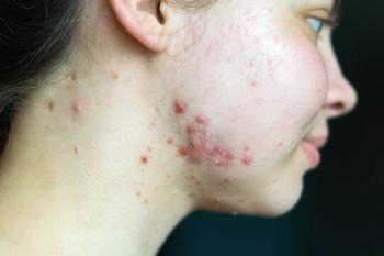
Venous ulcers are common on medial or lateral aspect of the ankle, in the malleolus area, associated with varicosities, surrounding haemosiderin and induration - lipodermatosclerosis. Venous ulcer location often distinguishes it from arterial ulcers of the lower extremity, which present anteriorly on the leg and somewhat more proximal, because they have less reduplication of arterial blood supply there. Atypical causes of wounds often present in unusual locations, for example, on the posterior aspect of the leg or dorsum of the foot. However even these ulcers may be complicated by insufficient arterial blood supply so assessing arterial supply in all lower extremity wounds is the first step.
Dr. Levine: How do we do that?
Dr. Kirsner: The simplest way is to palpate pulses and then perform an ankle brachial index, which is the systolic blood pressure in the ankle over the arm. When supine in healthy people they should be equivalent or a ration of 1. If you have diminished ankle brachial index, meaning the systolic pressure is lower, it correlates with worse arterial disease. A systolic pressure below about 0.8 should trigger a dermatologist to do three things:
- A dermatologist may want to refer to a vascular surgeon to determine if opportunities to improve blood flow surgically exist.
- A dermatologist might want to refer to the primary care doctor or cardiologist, because vascular disease in the legs is associated with vascular disease in the coronary and carotid arteries. So a low ABI in the lower extremity may be an indicator of cardiac or cerebral vascular disease.
- A dermatologist may wish to reduce the strength of the compression bandage prescribed, standard care for venous lucers, so not to restrict arterial blood flow.
Dr. Levine: Could you describe exactly how one does a blood pressure determination of the lower extremities?
Dr. Kirsner: Sure. It’s somewhat similar to the upper extremities. You place a cuff around the calf and inflate it to about 200 mm Hg, and then slowly release it. Then you are looking for the first return of arterial flow or pulse. You can do this in several ways.
- You can have a microphone or Doppler that you can listen to for the pulse;
- You could put your finger or hand over the area where the pulse would return, either the posterior malleolus or the dorsum of the foot;
- You could use the stethoscope
When you first hear the blood return, that’s the number you are looking for, that’s the systolic blood pressure. The ratio of that with the blood pressure in the arm would give you the ankle brachial index.
Dr. Levine: If you could feel somebody’s distal pulses, does that tell you something or is that not good enough?
Dr. Kirsner: It’s probably not good enough. Except in situations such as a young person who has a traumatic wound to his leg when checking pulses is probably all you need to do, for other patients studies have shown that palpating pulses is not reliable, due to its subjective nature. We have all been in the room where one person says I feel athe pulse ; the other person says I hardly feel it; and you don’t know where the truth lies. So obtaining objective measures is better.
Dr. Levine: Let’s talk about some of the agents that you use to treat leg ulcers. I know that a lot has changed in the last 150 years, but the Unna boot designed around the 1850s seems still to be a useful tool. How do you use Unna boots and other old fashioned remedies for leg ulcers?
Dr. Kirsner: There is no question that compression is the gold standard for venous ulcers and all lower extremity ulcers, if there is good arterial flow. The Unna boot, in its original form, would harden almost like a cast and typically, providing compression when the person was walking. When the person’s calf muscle would activate through walking, it would hit against that hard material and to reinforce the calf muscle.
For people with good arterial supply, there may be a slight benefit for using elastic compression as opposed to inelastic compression to speed healing, as it provides compression when a patient is walking and when they are not. The Unna boot plus an overlying elastic bandage transform the Unna boot from inelastic to elastic compression and is likely as good as any of the other systems that are available that have two, three, and four layers. However, it is known that multilayered elastic systems are better than just a single layer and elastic compression is better than inelastic.
However, if arterial disease is present, then inelastic compression is preferred so that you are not squeezing the limb, for example, when the person is supine in bed at night.
Dr. Levine: Could you discuss the role of surgical debridement of leg ulcers?
Dr. Kirsner: There is fairly good data for surgical debridement for diabetic foot ulcers and it’s considered the standard of care as I described earlier.
For venous leg ulcers, less data exists for debridement. Some studies have suggested it has been beneficial, others have found no benefit. As data is lacking, clinicians who debride, do it based on its rationale. That is they want to remove the bacteria or biofilms that are often in the base of a venous leg ulcer, remove any senescent or unresponsive cells within the wound bed, as well as unresponsive keratinocytes and fibrotic tissue around the edge.
Currently debridement is considered a two-phase approach. Initially, when feasible, an excisional debridement is performed and thereafter a maintenance or selective debridement is performed periodically.
The initial excisional debridement is performed following anesthesia. Using a scalpel to the wound is saucerized to remove cells and unhealthy tissue in the wound bed and wound edge. The wound will get bigger - slightly deeper and slightly wider. This allows healthy cells to migrate into the wound.
In subsequent visits, selective or maintenance debridement, is performed. Using a curette selectively chosen tissue within the wound bed that appear unhealthy, such as necrotic tissue or slough are removed.
Dr. Levine: There are number of so-called new technologies which have come on the stage over the last several years that it’s hard to understand the data whether they are helpful or not. Two that come to mind are Medihoney and some of these debriding enzymes; could you comment on those products?
Dr. Kirsner: There are typically five types of debridement.
- Surgical debridement or sharp debridement is most preferable if your patients can tolerate it.
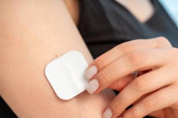
- Autolytic debridement uses an occlusive dressing to keep the patient’s own proteolytic enzymes in the wound area help to debride.
- Mechanical debridement, in which you apply and remove dry dressing, can be painful and is not optimal.
- Enzymatic debridement includes the enzymes, like collagenase to have the ability to debride by sloughing off dead tissue. Some people consider Medihoney under this category while others suggest it works an an autolytic debriding agent.
- Biological larval debridement involves the use of a specific type of maggot to dissolve dead tissue and disinfect the wound.
These are different tools to get to the same outcome. The major obstacle with using some of the enzymatic debriding agents is that these topical debridement agents were meant to be applied frequently. Typically when you treat a patient with a venous leg ulcer, you place a compression wrap on and you leave it on for up to a week or longer, depending on the amount of drainage. So, the enzymatic debriding tools don’t work the way that they should, because they are not being applied as frequently as they should be. Typically I relegate those enzymatic debriding agents to patients who aren’t getting weekly dressing changes but rather daily dressing changes, such as those patients in nursing homes.
Dr. Levine: My old mentor Gerald Lazarus, M.D., used to say that the ulcers have plenty of their own enzymes and adding them is not helpful. What is your view of that?
Dr. Kirsner: That’s exactly the concept behind autolytic debridement: You cover the wound and let the patients’ own wound proteases help deride the wound. Sometimes proteases are excessive and they can be destructive to the wound. So there is really a balance. New technologies are being developed to detect how much proteases are in the wound so that you can know if there is a healthy balance.
Dr. Levine: Could you comment on these sophisticated woundcare systems that people use with multiple agents applied in various ways.
Dr. Kirsner: Yes. There are two ways to think of systems. The first way is that there has been development of many wound centers throughout the country. There are probably about 1,500 wound centers often associated with hospitals; often with a multidisciplinary panel of physicians. A patient visits a setting in which they see physicians and nurses who have a special interest and expertise in wound healing and who follow evidence-based algorithms in their treatment. For many patients that’s beneficial, because most physicians may not have much knowledge about wounds.
The second idea of a "system" to treat wounds is based on why these wound centers were initially developed. They were initially developed to deliver something called platelet-derived wound healing formulaor Procurin. With this treatment, a patient has their blood taken and then platelets are separated and then activated. Activated platelets contain growth factors,which can be reapplied onto the wound. While it appears to be effective, other treatments may be equally or more effective.
For example, recombinant platelet-derived growth factors seem to be even better than platelet extracts. Perhaps it is because that not every patient’s platelet extract is the same. An elderly person may not have a good platelet extract or wound healing formula as a younger person, or it even varies day to day or week to week. There is currently not any nbiomarkers to know whether this treatment has consistent biologic activity. If that would be developed, then you can be more selective with this therapy.
Advertisement
Related Content
Advertisement


Latest CME


Advertisement
Advertisement
Trending on Dermatology Times
1
FDA Accepts Addition of Bemotrizinol as First New Sunscreen Ingredient in 20 Years
2
Dual-Action Microneedle Patch Shows Promise for Psoriasis in Preclinical Study
3
AI Analysis Reveals Menopausal Hair Loss Linked to Systemic Health and Inflammation
4
Novel Botanical Moisturizer Demonstrates Superior Efficacy Over Metronidazole in Phase 2 Rosacea Trial
5