
|Articles|September 24, 2014
Melanoma detection involves more than using ABCDs
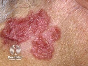
Melanomas can appear similar to other diagnoses, a fact that clinicians should keep in mind and why they should use tools like dermoscopy as an adjunct to making a diagnosis of melanoma, an expert says.
Advertisement
Melanomas can appear similar to other diagnoses, a fact that clinicians should keep in mind and why they should use tools such as dermoscopy as an adjunct to making a diagnosis of melanoma, according to an assistant professor at Harvard Medical School, Boston.
"Melanomas that don't follow the ABCDs can be difficult to diagnose, sometimes mimicking seborrheic keratoses, basal cell carcinomas, dermatofibromas, and even benign lentigines," Caroline C. Kim, M.D., F.A.A.D., who serves as director of the Pigmented Lesions Clinic at Beth Israel Deaconess Medical Center tells Dermatology Times. "The ABCDs are well-described clinical features of melanoma: Asymmetry (A), where one half of a mole is different than the other half, border irregularity (B), where the edges are jagged or uneven, color (C), where there are multiple colors to a mole (three or more), and diameter (D), where the diameter of the lesions exceeds 6 mm. The ABCDs are exemplified typically by the superficial spreading melanoma subtype; however, other subtypes, such as nodular and amelanotic melanomas may not exhibit these features.
“This is why we encourage utilizing clues from the clinical history such as change, and symptoms such as itching or bleeding of the lesion as well as clinical clues from dermoscopy as ways to improve our diagnostic accuracy,” Dr. Kim says. “Short-term follow-up is also a useful tool for borderline suspicious lesions if the patient is able to return for another visit.”
Dr. Kim says that a helpful guide for clinicians and patients alike for melanoma detection is the "ugly duckling" rule.
“This rule highlights a lesion that does not look like the rest of the patient’s typical mole pattern, which is a useful rule for complex, moley patients, and for melanomas that don’t fit the ABCD rule,” Dr. Kim says. “If there is a lesion that looks or acts differently from the result of the lesions on a patient for any reason, this is a lesion we should be looking at more carefully to consider a biopsy.”
Genetics and phenotypic factors, such as having fair skin, having red and light hair, having a tendency to freckle, exposure to significant amounts of ultraviolet radiation, and being immune suppressed increase the risk of developing melanoma, she says. Still, melanoma can occur in anyone, Dr. Kimsays.
"Every individual carries a certain risk for developing melanoma,” she says.
When melanoma does arise, it is more often not a case of family history of melanoma.
"Familial melanoma only comprises about 10 percent of melanomas," Dr. Kimsays. "It is more often the sporadic case that is a typical melanoma patient."
The incidence of melanoma is on the rise, and it's thought to be increasing by about 5 to 10 percent annually. According to the American Cancer Society, about 76,100 new melanomas will be diagnosed in 2014, and about 9,710 people are expected to die of melanoma in 2014.
The average age of a patient who is diagnosed with melanoma is 57, but it is commonly found in younger patients as well, even rarely in children, Dr. Kim says.
Technologies serve as aids in making the diagnosis of melanoma.
"Most dermatologists are using dermoscopy, which gives us in vivo view of the pigment pattern within the skin,” she says. “Studies have shown that the accuracy for detecting melanoma increases with the use of dermoscopy when the clinician is properly trained.”
Emerging aids in the diagnosis of melanoma include multispectral imaging and confocal microscopy, Dr. Kim says.
"There is a need for new technologies,” Dr. Kim says. “More study is needed to better understand how they may play a role in our clinical practices.”
Surgical excision remains the primary treatment for early stages of melanoma, with surgical margins determined by Breslow depth and other histologic features of melanoma.
The American Joint Committee on Cancer 2009 staging criteria outlines that thin melanomas of less than 1mm thickness with high-risk features, including ulceration or mitotic count of 1/mm2 or greater are considered T1B tumors. T1B tumors and those that are more advanced may be offered sentinel lymph node biopsy in many melanoma programs, Dr. Kim says.
In addition to standard surgical practices for melanoma management, clinicians should be aware of oncologic practices for advanced melanoma patients in their area, to consider referral of patients for adjuvant and advanced melanoma therapies, Dr. Kim says.
Disclosures: Dr. Kim had no financial disclosures.
Advertisement
Related Content
Advertisement


Latest CME


Advertisement
Advertisement
Trending on Dermatology Times
1
What Dermatologists Need to Know About the First New Sunscreen Ingredient in Decades
2
The Expanding Role of Immunotherapies for Non-Melanoma Skin Cancers
3
Once-Daily Zasocitinib Rivals Injectable Biologics for Skin Clearance, Phase 3 Data Show
4
Filling the HS Treatment Gap: Ruxolitinib Targets Early-Stage Disease
5