Mastocytosis: Be alert to vague symptoms
Loma Linda, Calif. — Dermatologists don't discuss mastocytosis much because it is rare and confusing, but, in large metropolitan areas, the incidence rate can add up to many patients, says Linda Golkar, M.D., assistant professor of dermatology, Loma Linda University, Loma Linda, Calif. Learning when to consider the disease, what diagnostic tests to perform and how to exclude involvement of other organs are required skills for front-line treatment.
"Often primary care physicians are not able to tie together the cutaneous presentation with other vague symptoms, such as itching, abdominal pain and diarrhea," she tells Dermatology Times. "I've seen patients who were misdiagnosed with congenital melanocytic nevi, insect bites and impetigo."
Dr. Golkar specializes in the treatment of children and currently has 14 pediatric cases of mastocytosis in her caseload. She defines the disease as a group of disorders originating from a proliferation of mast cells.
While the exact incidence is unknown, one British study estimated it at 1:150,000. Importantly, 55 percent of the time it begins before the age of 2 and 10 percent of the time between ages 2 and 15. Onset for the remaining 35percent occurs in the 20-to-40 age interval.
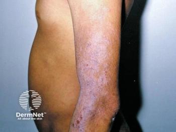
"In adults, the papules are tiny and reddish brown. They only gradually multiply in number," Dr. Golkar says. "With children, the papules are fewer but much larger in size. In both populations, they are concentrated on the trunk. Children tend to form bullae, but that's rare in adults."
Cutaneous mastocytosis in children usually resolves by adolescence. Systemic mastocytosis is mostly an adult phenomenon, sometimes progressive in course.
Receptor and mutation Mast cells originate in the bone marrow from CD34+ progenitor cells and circulate in the peripheral blood as precursor CD34+ cells, migrating into tissues where they differentiate and mature with help from growth factors, receptors and antigens.
"In the last 10 or so years, we've discovered that the c-kit receptor plays a central role in the biological development of mast cells and in the pathophysiology of adult onset disease," Dr. Golkar says. "A mutation at the codon 816 position leads to constant activation of the receptor and constant production of mast cells that collect in the skin."
The c-kit mutation suggests that mastocytosis is clonal in nature. Researchers regard systemic forms as myeloproliferative disorders.
Only children with persistent or progressive versions of the disease express the c-kit mutation, suggesting a different - and still unknown - pathophysiology for juvenile onset.
Diagnosis, treatment approach Dr. Golkar begins management with a history, asking if the patient has experienced flushing, pruritus, diarrhea or episodes of passing out, to determine the degree of mast cell load. Upon physical examination, she strokes papules to see if they urticate and palpates for lymphadenopathy, hepatomegaly and splenomegaly. She also performs a biopsy (histological evidence of increased mast cell proliferation establishes a diagnosis) and orders a complete blood count, liver tests and serum tryptase level (biochemical evidence is supportive).
"A serum tryptase over 20 ng/ml suggests systemic mastocytosis," she says.
The goal in treating cutaneous mastocytosis is to reduce symptoms and improve the patient's cosmetic appearance. Treatment should be conservative.
Newsletter
Like what you’re reading? Subscribe to Dermatology Times for weekly updates on therapies, innovations, and real-world practice tips.


