Laser options grow for hypertrophic, keloid scarring
Revolutionized by the advent of fractional laser technology, laser surgery for scars continues to evolve. However, clinicians continue to be confronted with a wide variety of scar types without a well-established treatment algorithm.
Dr. Kaufman
Dr. Green
Among the most common types of scars that laser surgeons encounter are acne scars, hypertrophic scars, keloids, burn scars, postsurgical scars, hyper- and hypopigmented scars and atrophic scars. Choosing the ideal device, parameters and treatment intervals can be quite challenging.
Current lasers indicated for the treatment of scars include the ablative (CO2, Er:YAG, Er:YSGG) and nonablative (1,540 nm/1,550 nm erbium glass) fractional resurfacing devices; potassium titanyl phosphate (KTP) laser; and the intermediate-pulsed and Q-switched Nd:YAG lasers. Choosing the proper laser for each different type of scar is imperative, as worsening of scars can also occur with laser treatment.
In fact, Apfelberg and colleagues first described argon laser treatment of hypertrophic scars (Apfelberg DB, Maser MR, Lash H, et al. Lasers Surg Med. 1984;4(3):283-290). Further investigations revealed that the argon laser itself could induce scarring, and the pulsed dye laser (PDL) was subsequently utilized to treat argon-laser induced scars (Alster TS, Kurban AK, Grove GL, et al. Lasers Surg Med. 1993:13(3):368-373).
In this month’s column, we will discuss laser treatment for hypertrophic scars and keloids.
Pulsed dye laser
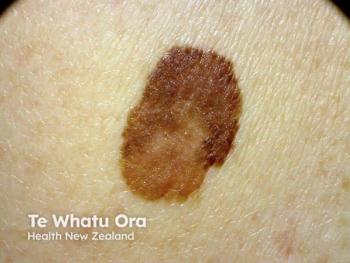
Hypertrophic scars and keloids are often grouped together in discussions of abnormal scarring, but their histology and pathophysiology do differ. As we dermatologists know, hypertrophic scars clinically do not extend beyond the margin of the original scar or injury, whereas keloids extend beyond these borders. Additionally, the histology of hypertrophic scars reveals vascularity, which is generally absent in well-formed keloid scars.
The increased hemoglobin chromophore in hypertrophic scars suggests that these scars would be more amenable to treatment with PDL if one assumes that the primary mechanism of action is the treatment of the vascular component of the scar. In fact, the most commonly reported laser for treatment of hypertrophic scars and keloids is the pulsed dye laser. But there are other proposed mechanisms of action.
In a well-designed controlled trial, Yang et al demonstrated that connective tissue growth factor is significantly downregulated in keloids after three PDL treatments (Yang Q, Ma Y, Zhu R, et al. Lasers Surg Med. 2012;44(5):377-383). Transforming growth factor beta expression in keloid tissue was also reduced after multiple treatments with the PDL (Kuo Y-R, Jeng S-F, Wang F-S, et al. Laser Surg Med. 2004;34(2):104-108).
The same group found a concomitant increase in matrix metalloproteinase 13 with PDL treatment (Kuo Y-R, Wu W-S, Jeng S-F, et al. Lasers Surg Med. 2005;36(1):31-37). If these changes in cytokine expression correlate with the clinical improvement of PDL-treated scars, then it would be more acceptable to group hypertrophic scars and keloids into a single category, as these laser-induced cytokine changes should be observed in both forms of exuberant scar formation.
This cytokine expression profile would not explain the efficacy of PDL for other forms of scarring, such as erythematous scars or striae distensae, as these are most likely based on selective photothermolysis of the target chromophore hemoglobin.
Addressing the variables
Hypertrophic and keloid scars can be treated effectively with PDL, but there are many variables that can affect treatment outcomes. The literature supports the idea that multiple sessions are needed for optimal treatment or prevention of hypertrophic scarring.
One study noted no difference in fresh surgical scars receiving a single treatment with PDL at the time of suture removal (Alam M, Pon K, Van LaBorde S, et al. Dermatol Surg. 2006; 32(1):21-25). In addition, short pulse widths (0.45ms) seem to offer better outcomes than long pulse widths (40ms) (Manuskiatti W, Wanitphakdeedecha R, Fitzpatrick RE. Dermatol Surg. 2007;33(2):152-161). The results of PDL treatments with a 0.45 ms pulse width did not affect the clinical result when compared to 1.5 ms pulse treatments (Nouri K, Elsaie ML, Vejjabhinanta V, et al. Lasers Med Sci. 2010;25(1):121-126), however.
Several studies have shown improvement in pruritus, skin surface texture, scar height and erythema after monthly treatments with PDL with short pulse widths and medium to low fluences. (Alster TS, Williams CM. Lancet. 1995;345(8959:1198-1200; Kono T, Erçöçen AR, Hiroaki N, et al. Ann Plast Surg. 2003;51(4):366-371; Manuskiatti W, Wanitphakdeedecha R, Fitzpatrick RE. Dermatol Surg. 2007;33(2):152-161).
As for the use of PDL to facilitate normal postsurgical healing, the data is contradictory. Some studies suggest the ability to prevent scarring with multiple sessions of PDL beginning on the day of suture removal (Conologue TD, Norwood C. Dermatol Surg. 2006;32(1):13-20; Nouri K, Jimenez GP, Harrison-Balestra C, Elgart GW. Dermatol Surg. 2003;29(1):65-73), whereas others did not find a significant difference between immediate and delayed PDL treatments (Chan HH, Wong DSY, Ho WS, et al. Dermatol Surg. 2004;30(7):987-994).
The 532 nm KTP laser as well as the intermediate-pulsed Nd:YAG (non-contact) have also been reported to improve the appearance of hypertrophic scars with multiple sessions (Cassuto DA, Scrimali L, Sirago P. J Cosmet Laser Ther. 2010;12(1):32-37; Akaishi S, Kaoke S, Dohi T, et al. Eplasty. 2012;e1).
Other options
Nonablative fractional devices play a role in the treatment of scarring. One group used the 1,550 nm erbium glass fractional laser and found an improvement in hypertrophic scars (Niwa ABM, Mello APF, Torezan LA, Osório N. Dermatol Surg. 2009;35(5):773-778). However, a study using a 1,320 nm/1,440 nm nonablative fractional laser showed minimal improvement after several treatment sessions (Babilas P, Schreml S, Eames T, et al. Lasers Med Sci. 2011;26(4):473-479).
Carbon dioxide lasers have also been reported for the treatment of hypertrophic scars and keloids. Older studies describe high recurrence rates, however, thereby mitigating the usefulness of the full-field CO2 laser.
Newer fractional devices have improved outcomes dramatically. Current literature demonstrates good results with fractional CO2 for burn scars. The use of fractional CO2 in hypertrophic scars and keloids of other etiologies is not well described.
Clinical scar quality of hypertrophic scars and keloids can be improved with several sessions of short-pulsed PDL. The data is less clear regarding postoperative scar prevention with lasers, but if attempted, treatments described in the literature start on the day of suture removal and require numerous sessions.
Other vascular lasers, such as the 532 nm KTP and the 1,064 nm Nd:YAG, have also been shown to be effective. The fractional devices, both nonablative and ablative, are rapidly gaining a foothold in this treatment category as well.
The authors prefer to treat erythematous hypertrophic scars and keloids with PDL or KTP lasers with short pulse durations followed by nonablative fractional
1,540 nm or 1,550 nm laser, and intralesional Kenalog (triamcinolone acetonide, Bristol-Myers Squibb) concentration titrated based on scar thickness) in the same session. We look forward to more standardized future studies using fractional devices for these types of scars to offer clinicians additional guidance. DT
Newsletter
Like what you’re reading? Subscribe to Dermatology Times for weekly updates on therapies, innovations, and real-world practice tips.


