Laser complications: Even experienced operators can incur negative reactions
We have discussed lasers, and some of basics of lasers, and now I would like to take a break from some of the science to present examples of the more clinical side of lasers.
Key Points
Laser complications can occur even in the best of hands. At the University of Miami, we are being referred an increasing number of laser complications, most likely due to the increased use of these devices and, often, the lack of supervision and training of operators.
It is important to remember that complications from laser treatment can happen to even the best-trained operator, but obviously, increase with lack of experience.
Case study
A female patient presented to the University of Miami with complaints of white spots on her chest after a laser procedure two years earlier. She went for a laser treatment to because her chest was red and "blotchy." She reports having two sessions done with a laser in a clinic.
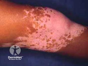
On physical examination, the patient had depigmented macules and atrophic plaques in a checkerboard pattern on her chest. Each scar measured approximately 5 mm to 7 mm in diameter.
The untreated areas were erythematous and blanched on diascopy. Other areas were slightly hypertrophic and depigmented.
According to the patient's records, the patient was treated with a pulsed dye laser with cryogen cooling. The first treatment was done with conservative settings. Settings for the second treatment were not recorded in notes.
Discussion
From the history, it is clear that this patient presented for treatment of poikiloderma. Treatment with a pulsed dye laser for this condition is a reasonable choice; however, the patient ended up with a poor outcome.
The pulsed dye lasers are commonly used for this condition, whether it be on the face, neck or chest area. Wavelengths of 585 nm and 595 nm, common with the pulsed dye lasers, use hemoglobin as the target chromophore, ablating the visible vessels seen in poikiloderma.
If there is a pigment component to the poikiloderma, it will need to be treated in another manner, usually with a combination of topical agents, such as hydroquinone or retinoids, or a different laser/light system.
Other laser and light choices for poikiloderma include intense pulsed light (IPL) devices, KTP lasers and nonablative fractional resurfacing. These devices are all able to produce successful results on poikiloderma when used with the proper parameters.
When operating a pulsed dye laser in sensitive areas or on patients with darker skin types, care should be taken to provide adequate cooling. Malfunction of a cooling apparatus can lead to blistering and scarring of the skin.
Shorter pulse widths - with all the energy delivered over a very short period of time - can also lead to blistering.
Newsletter
Like what you’re reading? Subscribe to Dermatology Times for weekly updates on therapies, innovations, and real-world practice tips.


