

|Articles|July 8, 2020
How to Safely Treat Patients with Darker Skin Types
Dr. Suzan Obagi says she’s not afraid of causing PIH in darker skin types. She offers her expertise and approach to understanding and treating pigmentation issues.
Advertisement
It is not unusual for me to see a patient in consultation for skin concerns only to find out that they were referred by their dermatologist or cosmetic surgeon because of fear of treating their darker skin complexion without risking more pigmentation issues. While I am thankful that the physician referred the patient if they were concerned about treating their skin safely, it makes me wonder how many patients out there are being turned away from treatments based solely upon their skin color.
I think we have been taught, rightfully so, to be concerned about post-inflammatory hyperpigmentation (PIH) when treating darker-skinned patients, but there are steps we can take to prepare the patient’s skin well so that we can safely treat them.
First, let’s look at the difference between ephelides (freckles), lentigines (sunspots), PIH, and melasma. Patients may have a genetic predisposition to forming ephelides, but that gene varies racially. Biopsies show increased pigmentation but not increased melanocytes; however, ephelides have a predilection for sun-exposed skin. This suggests that there is an overproduction of melanin by melanocytes. Solar lentigines, on the other hand, arise in areas where repeated sun exposure induces mutations, resulting in increased melanin production and abnormal pigment retention by the keratinocytes. In these lesions, there is a mild increased number of melanocytes as well as evidence of photodamage in the dermis.
PIH is a temporary overproduction of pigment in response to an epidermal injury or trauma. Usually, this injury is thermal but PIH can be seen in patients that excoriate their skin or that have had inflammatory lesions in the skin. Rarely, severe inflammation can cause melanin to drop into the dermis and to be engulfed by melanophages. PIH is worsened by sun exposure.
Melasma, on the other hand, is a much more complex disorder that may have a genetic predisposition. Melasma arises mostly in women of child-bearing age but it can be seen in men and older women. In melasma, some unknown trigger has induced melanocytes to overproduce and distribute melanin to the keratinocytes. Sometimes, this pigment can drop into the dermis and be taken up by melanophages resulting in dermal melasma. While the trigger may be elusive, there is certainly a link to things that worsen melasma. These include ultraviolet light and certain visible light wavelengths, hormones (pregnancy, oral contraceptives, hormone IUD), and certain topical agents (chemical sunscreens, fragrances and certain botanical agents), heat (spas, saunas, hot yoga, cooking). Once these melanocytes are sensitized and over-produce pigmentation, it is challenging to quiet them down or to clear the melasma fully.
Evaluation & Diagnosis
When I see patients with dyschromias, it is important to assess the following:
- Diagnosis of condition: ephelides, lentigines, PIH, melasma
- Is this strictly epidermal or is there a dermal component?
- What has been tried, what has worked and what has failed or worsened the condition?
- For melasma, is the patient exposed to exogenous hormones such as oral contraceptives, excessive soy intake or the presence of a hormone IUD?
- How motivated the patient is to follow an aggressive skincare regimen?
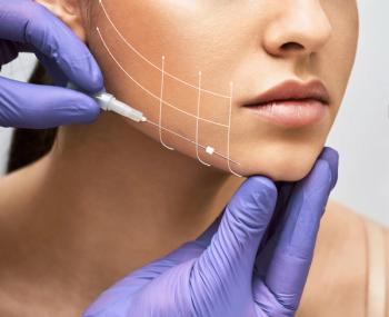
Patients with ephelides and lentigines can be safely treated with light peels and non-ablative fractionated lasers, Q-switched lasers, and broadband light (BBL). However, the darker the patient’s complexion, the longer I pretreat with a comprehensive skincare regimen before performing any of these treatments. The risk of not doing so is that you will induce PIH during the healing process.
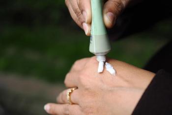
Patients with PIH — the dreaded complication — are actually easy to treat. It may resolve with proper management in one month, three months or six months, but in the end, it all resolves. Patients that develop hyperpigmentation in response to acne or oven burns are at risk for PIH from any face procedure. PIH is not permanent thus it does not scare me if it arises, but I try my utmost best to minimize the risk of PIH and to treat it preemptively. Any patient at risk for PIH should be pretreated with a comprehensive skincare regimen, and once they have healed after a procedure, the patient should quickly resume the topical regimen. If, despite this, PIH develops, the patient must be diagnosed quickly and started on a series of 30% salicylic acid peels at two- to four-week intervals until the PIH resolves. Being more aggressive with peels, or by performing a laser (heat trauma), will only aggravate the condition. Lasers have no role to play in treating PIH.
Melasma is the one condition for which I do not over-promise the results. If the patient has eliminated all their contributing factors (hormones, heat, certain topical agents) and they have used their comprehensive skincare regimen faithfully, epidermal melasma should show considerable improvement by six weeks (one skin cell turnover cycle). If at six weeks the patient is doing well, I then see them back at three months. However, if at six weeks, they are not improved despite using their products correctly, I add light peels to their regimen with 30% salicylic acid every four weeks. At three months, I reassess their skin. If it is considerably improved, I continue the skincare regimen unchanged for one year. The light peels can continue. If the patient is only moderately improved, I might add a non-ablative fractionated laser combined with a salicylic acid peel or Jessner’s solution peel. However, if there is little change at the three-month evaluation, it may indicate that the pigmentation is dermal. If so, I may proceed with a medium-depth trichloroacetic acid (TCA) peel to try to eliminate some of the dermal melanophages. I do not use more than a 24% or 25% concentration TCA peel, as higher concentrations can cause an inflammatory reaction in the skin. Of course, once the patient has healed, they resume their topical skincare regimen immediately. It is important not to let the melasma rebound.
Advertisement
Related Content
Advertisement

Latest CME


Advertisement
Advertisement
Trending on Dermatology Times
1
Switching from Biologics to Upadacitinib Yields Progressive Improvements in Moderate to Severe AD
2
Sunscreen’s New Blind Spot: Fashion Trends, Tanning Myths, and the Rise of Gluteal Melanomas
3
New Consensus Defines Remission, Disease Activity in Atopic Dermatitis
4
Derm Dispatch: New ICD-10 Code Brings Validation to Patients With Topical Steroid Withdrawal
5