
|Articles|May 1, 2013
Facial dermatitis may mask other conditions
The archetypal red face of dermatitis may conceal more diagnoses than originally meet the eye, according to an expert who spoke at the annual meeting of the American Academy of Dermatology.
Advertisement
Miami Beach, Fla. - The archetypal red face of dermatitis may conceal more diagnoses than originally meet the eye, according to an expert who spoke at the annual meeting of the American Academy of Dermatology.
When a patient presents with a red face, “What we see may not necessarily be a single diagnosis. We must consider multiple diagnoses,” says Kalman L. Watsky, M.D., clinical professor of dermatology, Yale University School of Medicine, New Haven, Conn. Conditions that commonly present with facial erythema can include actinic erythema, rosacea, allergic contact dermatitis and seborrheic dermatitis, he says.
Regarding rosacea, he says, dermatologists have little trouble identifying the characteristic papulopustular, inflammatory type.
“But patients with rosacea also may present with just telangiectasia (erythematotelangiectatic rosacea/ETR), which can be difficult to distinguish from telangectasias related to chronic sun exposure (telangiectatic photoaging),” Dr. Watsky says.
In this regard, Yolanda Helfrich, M.D., assistant professor of dermatology, University of Michigan, Ann Arbor, has presented research suggesting that mast cells and neuropeptide expression are increased in ETR in comparison to telangiectatic photoaging (Helfrich YR, Varani J, Fisher G, et al. J Invest Dermatol. 2012;132:S1-S18. Abstract 093).
Clinically, Dr. Watsky says, Dr. Helfrich’s research demonstrates that in ETR, the erythema occurs more on the central face, versus a more lateral presentation in telangiectatic photoaging.
“Patients who complain of sensitive skin and/or symptomatic flushing are more likely to have rosacea,” Dr. Watsky says.
Presently, he says that for some cases of ETR, “The only effective treatments are vascular lasers.” Additionally, among treatments under development, a study in ETR has shown that once-daily treatment with topical brimonidine tartrate gel 0.5 percent for four weeks achieved statistically superior results versus vehicle within 12 hours on treatment days one, 15 and 29 (all P<0.001; Fowler J, Jarratt M, Moore A, et al. Br J Dermatol. 2012;166(3):633-641).
Additional conditions
Patients with rosacea also may have coexisting seborrheic dermatitis, Dr. Watsky says.
“So on a given day, they may present with features that are more consistent with seborrheic dermatitis than rosacea,” he says. If a doctor overlooks the rosacea and treats the seborrheic dermatitis with topical steroids, he warns, these will exacerbate the rosacea.
Additionally, “Be on the lookout for ocular rosacea. Patients with ocular rosacea will typically complain of a foreign-body sensation in the eye. They generally respond well to oral antibiotics.”
“A characteristic appearance and histology can help,” in diagnosing facial dermatitis via biopsy, Dr. Watsky says. “However, a biopsy won’t help you make a definitive diagnosis.”
To that end, “Look at the whole patient and see if there’s evidence for dermatitis in other locations. Also, remember that dermatitis may be multifactorial - for example, a patient with facial dermatitis may have underlying seborrheic dermatitis, and also may have developed an allergy to some topical or airborne contactant. In such cases, patch testing provides an objective tool” to distinguish between these entities.
Generally, he says, “Patch testing with the Food and Drug Administration-approved series of allergens is a reasonable screening tool. But it often misses important allergens,” particularly in facial locations.
If this happens, “We then test with an expanded series of allergens, also considering the patient’s exposures.” If the patient uses certain hair dyes, hair treatments or other personal care products, for example, Dr. Watsky patch tests against these types of allergens.
“If the allergy seems work-related, it’s very important to consider airborne exposures in particular.” Such tests can tell the patient what materials to avoid, if possible.
Maximizing efficacy
“Even after you’ve done your evaluation, you may not find a specific etiology. It’s something that the patient likely will have to cope with lifelong. For example, we don’t cure atopic dermatitis - it requires long-term management, with an eye toward maximizing safety and efficacy.”
In such cases, he says, treatments for facial dermatitis largely mirror those of dermatitis in general.
“But some special areas need to be addressed. In particular, people with atopic dermatitis or seborrheic dermatitis commonly have eyelid dermatitis. For people with chronic conditions who need ongoing therapy, if topical steroids don’t work well enough that you can use them sparingly, look for steroid alternatives such as tacrolimus and pimecrolimus,” Dr. Watsky says.
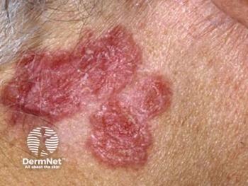
Conversely, he says that as with rosacea, dermatologists should not treat the redness and scaling of periorificial dermatitis with anti-inflammatory agents such as steroids. Clinically, he adds, “Periorificial dermatitis looks much like perioral dermatitis. But it occurs around the eyes or nose. You also may see tiny bumps with erythema and a bit of scale.”
Dr. Watsky typically treats periorificial dermatitis with low-dose doxycycline, or topical metronidazole or azelaic acid (though the latter can cause irritation).
“If there’s a significant inflammatory component, I also will use anti-inflammatories such as tacrolimus and pimecrolimus off-label,” he says.
Disclosures: Dr. Watsky reports no relevant financial interests.
Advertisement
Related Content
Advertisement


Latest CME


Advertisement
Advertisement
Trending on Dermatology Times
1
KT-621 in Atopic Dermatitis: What Early EASI and Pruritus Data Show
2
What Dermatologists Need to Know About the First New Sunscreen Ingredient in Decades
3
Filling the HS Treatment Gap: Ruxolitinib Targets Early-Stage Disease
4
Once-Daily Zasocitinib Rivals Injectable Biologics for Skin Clearance, Phase 3 Data Show
5