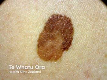
Evaluating suspicious lesions requires equipment, expertise
When analyzing suspicious lesions, any diagnostic tool is only as good as the physician using it. Besides patients' suspicions, dermatologists can rely on objective diagnostic tools including dermoscopy and total body photography.
Key Points
Besides patients' suspicions, dermatologists can rely on objective diagnostic tools including dermoscopy and total body photography, says James M. Grichnik, M.D., Ph.D., director, Melanoma Program, Sylvester Comprehensive Cancer Center; and professor, department of dermatology, University of Miami.
With dermoscopy, physicians can follow selected lesions over time using detailed baseline images, he says. In one study, investigators used dermoscopy to follow 318 suspicious lesions and found that over three months, 61 lesions had changed. These 61 lesions included seven melanomas - five in situ and two invasive. Overall, dermoscopy achieved a "hit" ratio of one melanoma per 7.7 biopsies (Menzies SW, Gutenev A, Avramidis M, et al. Arch Dermatol. 2001;137(12):1583-1589).
Total body photography allows physicians to compare moles' appearance on follow-up visits to how they looked in previous visits, Dr. Grichnik says. "The trick here is knowing who is at high risk. In general, those of us who obtain total body photos do so with patients who have multiple dysplastic nevi, particularly patients with a personal or family history of melanoma," as well as those who have large congenital nevi.
"If you see a lesion you are worried is melanoma, you should remove it immediately, at the initial visit. Thereafter, theoretically you shouldn't remove lesions unless digital photographs document a change" that causes concern, he says. "In our early studies, we found that when we used digital photos, the biopsy rate was around 0.25 per patient per year. One of 10 biopsied lesions was melanoma, and one out of four were melanoma or severe atypical nevi (Lucas CR, Sanders LL, Murray JC, et al. J Am Acad Dermatol. 2003;48(5):663-671)."
Making decisions
In deciding which changing nevi to excise, it's important to consider that "all nevi can change, and if you follow patients, somewhere on the order of 5 percent of lesions will change per year. Therefore, change itself - while important for melanoma diagnosis - is not sufficient reason to remove a lesion," Dr. Grichnik says. Only about 2 to 3 percent of changing lesions are actually melanoma, he says.
Patient age can provide guidance regarding which changing lesions to excise, he says. Specifically, the 5 percent of lesions that change annually masks, to a degree, the fact that patients under age 20 usually have a higher number of changing lesions (between 12 and 57 percent in one study), while older patients experienced far fewer changing lesions (Kittler H, Seltenheim M, Dawid M, et al. Arch Dermatol. 2000;136(3):316-320). Older patients also experienced fewer new moles, "and the number of melanomas increases with age," Dr. Grichnik says.
Additional research involving 1,862 lesions followed for an average of more than a year has shown that most benign nevi enlarged symmetrically, while the eight melanomas detected exhibited focal enlargement and change of shape (Kittler H, Pehamberger H, Wolff K, Binder M. J Am Acad Dermatol. 2000;43(3):467-476).
Somewhat similarly, Dr. Grichnik says that in the dermoscopy study that he and his colleagues conducted, "There were 16 melanomas. Five originated from spots that may have been small nevi; four originated from areas where we could find a small focal area of pigment; seven came from what appeared to be totally normal skin, and zero developed from obvious dysplastic nevi (Lucas CR, Sanders LL, Murray JC, et al. J Am Acad Dermatol. 2003;48(5):663-671)." These data reinforce the importance of considering the whole patient rather than following only a handful of suspicious nevi, he says.
"All the invasive melanomas, and 75 percent of melanomas overall, were thought to be nonuniform and changed. But it's interesting that about 25 percent of the melanomas included an in situ component thought to be uniform. So we need to be careful - some of the in situ melanomas in the early part of their growth may still be relatively uniform-appearing lesions," Dr. Grichnik says.
Newsletter
Like what you’re reading? Subscribe to Dermatology Times for weekly updates on therapies, innovations, and real-world practice tips.


