- Vol. 39 No. 05
- Volume 39
- Issue 5
Difficult to treat actinic keratoses responds to topical treatment with laser
Lasers might allow the topical treatment of skin cancers with chemotherapy combinations that are currently given systemically, researchers say.
"The biggest advantage would be reducing side effects when you go from systemic to topical treatment," said Merete Haedersdal, M.D., Ph.D., a dermatology professor at the University of Copenhagen, Denmark.
She described the innovation in April at the American Society for Laser Medicine and Surgery annual conference in Dallas.
Experiments in pigs have proven that creating arrays of holes with ablative fractional lasers can significantly accelerate topical uptake of drugs normally used systemically to treat squamous and basal cell carcinomas.
If successful in humans, the technique would mark a new chapter in the evolution of a laser technique that has already scored remarkable successes in other skin conditions, said Dr. Haedersdal, who has devoted the past 10 years to exploring its potential.
"The technique is taking off all over the world," she said. It has been tested in clinical trials on neoplastic lesions, photodamaged skin, scars, onychomycosis, alopecia and topical anesthetics, with the best evidence for actinic keratoses.
Fractional laser-assisted drug delivery most commonly uses erbium-doped yttrium-aluminum-garnet or carbon dioxide lasers operating at infrared wavelengths, with water as the target chromophore. Evaporating tissue, the lasers leave a matrix of vertical channels surrounded by a rim of coagulated tissue. The channels penetrate the stratum corneum, providing direct access to viable skin.
The patient experiences little discomfort, though topical anesthetics are sometimes used, she said.
"What we have been working on is establishing laser parameters, what kind of drugs can get in, these drug interactions and basic understandings," she said. The researchers are learning to vary their technique depending on the drug they want to deliver. Smaller molecules are absorbed more easily than bigger ones. Hydrophilic drugs are absorbed more easily than lipophilic drugs.
"We do know that not many holes are required - about 5% density. It's very subtle. If you crank up the laser you have the risk of systemic absorption. We want to keep it in the skin,” Dr. Haedersdal said.
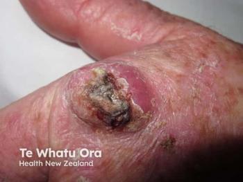
The technique has shown good success in treating actinic keratoses that are difficult to treat surgically because they cover large areas. The laser treatment enhances the uptake of a photosensitizer used as part of photodynamic therapy. "Before they might need 2-5 treatments, now with 1-2 treatments they achieve the same outcomes," she said.
Atrophic and hypertrophic scars
For atrophic and hypertrophic scars, the lasers enhance the uptake of corticosteroids or 5-ï¬uorouracil.
Now the researchers are hoping to apply the technique to cancer. "The paradigm shift is to move treatments from systemic treatments to topical treatments," she said.
The researchers have focused on non-melanoma skin cancers because they are so common, with 5.4 million cases in the United States alone.
Surgical resection or destruction can cause scars and disfigurement and may not fully remove tumors. Topical treatments have been limited by the poor penetration of the drugs into the skin. Already Dr. Haedersdal and her colleagues have achieved some success in a clinical trial treating basal cell carcinoma with photodynamic therapy.
Topical 5% 5-FU
In work led by Emily Wenande, MD, of Harvard Medical School in Boston, they are planning a trial using cisplatin and 5-ï¬uorouracil (5-FU). "If you combine the two drugs, it has a synergistic response in cell turnover," she said. The drug combination is currently used systemically to treat malignancies, including head and neck squamous cell carcinoma.
Topical 5% 5-FU is approved for superficial basal cell carcinoma but does not adequately clear deep lesions. Cisplatin's cutaneous uptake is so poor that it is currently limited to systemic therapy for metastatic nonmelanoma skin cancers.
To test the approach in the skin of live pigs, the researchers used a CO2 laser (10,600 nm) to generate channels in test sites, 2 x 2 cm. They applied customized wells made from wound dressings to these test sites and to other test sites on the pigs that were not treated by laser.
The test sites each received 1 of 9 interventions: cisplatin on intact skin, laser followed by cisplatin solution (30 or 60 minutes exposure), laser followed by cisplatin (30 or 60 minutes) and 5-FU cream for 5 days, 5-FU on intact skin, laser followed by 5-FU for 5 days, laser alone, and no treatment.
In the intact skin, the cisplatin remained mostly on the superficial skin layer. It reached peak concentrations between 2 and 5 hours of 20.03 µg/cm3 at 500µm depth and 5.09 µg/cm3 at 1500 µm depth. In the laser-treated skin cisplatin reached peak concentrations of 75.72 µg/cm3 at 500µm depth and 29.30 µg/cm3 at 1500µm depth. The differences were statistically significant (P < 0.005)
And the cisplatin concentrations in the laser-treated skin remained higher than in the intact skin, culminating in 16-26-fold greater deep deposition at 120 hours.
In the intact skin, no 5-FU was detected in the1500 µm layer until 120 hours. At that point, it reached 17.67 µg/cm3. By contrast, in the laser treated skin, 5-FU concentration reached 318.74 µg/cm3 at 1500µm depth after only 1 hour.
Between 48 and 120 hours, the laser's effects on the uptake of 5-FU had waned, such that there was no difference between the intact and laser-treated skin at 1500 µm. And at the more superficial layer, 5-FU concentrations were higher in the intact skin at 120 hours.
The researchers did not detect either drug in systemic circulation. And they did not find any difference in local skin reactions among the test sites, with negligible erythema and edema.
The skin treated with laser and either of the drugs showed more signs of inflammation, cellular apoptosis and, for 5-FU, necrosis of the epidermis. The increased apoptosis and immune cell infiltration, and reduced epidermal proliferation, was particularly marked in skin treated with both drugs after the laser. This suggested a synergistic, or at least additive, effect of the 2 drugs.
"We are much encouraged that we can develop a paradigm shift in dermo-oncology," said Dr. Haedersdal. She and her colleagues expect to begin a clinical trial by early summer. They are planning to use optical coherence tomography and confocal microscopy to measure the effects on tumors.
The approach could work with melanoma as well, but would require different drugs, she said.
Articles in this issue
over 7 years ago
Minimizing the risk of lidocaine toxicityover 7 years ago
Clinical variations in pyoderma gangrenosumover 7 years ago
Immune modulators offer promising outcomesover 7 years ago
Improving continence with laser treatmentsover 7 years ago
Can I be sued for off-label use within standard of care?over 7 years ago
Vertical dermatology integration without dermatologistsover 7 years ago
Key steps to avoid a malpractice lawsuitover 7 years ago
How to pair energy-based devices with cosmetic injectablesalmost 8 years ago
A single drug may control skin cancer treatment costsalmost 8 years ago
How deep an SCC lies within the skin correlates to lesion lengthNewsletter
Like what you’re reading? Subscribe to Dermatology Times for weekly updates on therapies, innovations, and real-world practice tips.

