
|Articles|July 7, 2014
Cutaneous drug reaction diagnoses require art, urgency
Successfully managing cutaneous drug reactions requires including drug reactions in the differential diagnosis of most cutaneous eruptions, and recognizing severe reactions where treatment may improve outcomes, an expert says.
Advertisement
Denver - Successfully managing cutaneous drug reactions requires including drug reactions in the differential diagnosis of most cutaneous eruptions, and recognizing severe reactions where treatment may improve outcomes, an expert says.
“Diagnosing cutaneous drug reactions is almost an art,” says David R. Adams, M.D., Pharm.D., professor of dermatology at Penn State Hershey Medical Center, Hershey, Pennsylvania. “Usually it’s based on the clinical appearance of the reaction and evaluation of the timing of medication administration. This can vary, depending on the type of reaction.”
Although biopsy and lab tests can help, he says, “The gold standard in diagnosing drug reactions is rechallenge. But we rarely ever do this, and clearly not intentionally in severe reactions.”
Rare but severe reactions
Among severe cutaneous drug reactions, he says, toxic epidermal necrolysis (TEN) manifests as erythroderma and painful desquamation of the skin. Similar symptoms characterize Stevens-Johnson syndrome, Dr. Adams says, although it affects 10 percent or less of the body surface area (BSA), including the epidermis and mucosa. Stevens-Johnson syndrome (SJS)/TEN overlap impacts 10 to 30 percent BSA, and TEN affects more than 30 percent BSA, he says.
“Mortality increases with increasing skin involvement, and with comorbidities such as HIV. TEN probably has a combination metabolic and immunologic pathogenesis. The key here is that if the patient’s skin hurts, there’s a very short window to make the diagnosis and begin treatment. TEN quickly progresses over several days, and these patients are at high risk for systemic complications.”
Along with discontinuing the possible associated drugs, he says, treating TEN requires referring the patient to a burn unit.
“Supportive care by the intensivist is important; avoid adhesives on the skin, a common nursing problem,” Dr. Adams says. “Ophthalmology needs to be involved to help manage ocular manifestations, which can lead to blindness.”
Unfortunately, he adds, the rarity of TEN makes it difficult to find strong studies that can help guide treatment.
“Depending on where you trained and your evaluation of current literature, some physicians use corticosteroids; some use IVIG or even cyclosporine. Some don’t use any medication other than supportive care. The important point is that after a couple days, none of these drugs are likely to have any impact because skin necrosis has already occurred,” Dr. Adams says.
Identifying DRESS syndrome
Patients experiencing drug reaction with eosinophilia and systemic symptoms (DRESS syndrome) always have a fever and some type of rash, often erythroderma, and varying degress of organ dysfunction, he says. Other symptoms can include facial edema and difficulty swallowing, he adds. Several dozen agents, including anticonvulsants, allopurinol and sulfonamides, have been associated with DRESS, Dr. Adams says.
“Systemic steroids often halt the process rather quickly, but you must give enough: methylprednisolone IV given at least 1 mg/kg body weight every 12 hours for several days, then transitioning to oral prednisone tapering over several weeks to months, depending upon response,” he says.
Nephrogenic systemic fibrosis (previously called nephrogenic fibrosing dermopathy) - a reaction to gadolinium-enhanced MRI in patients with advanced kidney disease - presents with very indurated skin that can make it difficult for patients to move their fingers or other extremities, Dr. Adams says. Other signs include hyperpigmented, hardened plaques. Although a flurry of cases occurred when this MRI contrast agent was first marketed in 1997, he says, newer radiology screening guidelines have reduced the use of gadolinium in the high-risk population with renal impairment. Because no treatments have been shown to consistently reverse the condition, he says, prevention is paramount.
Other drug reactions include acute generalized exanthematous pustulosis (AGEP). In one case, a resident was called to an ER consult regarding a patient diagnosed with a potential groin infection. Closer examination revealed a symmetrical pattern of small studded pustules in the groin area. AGEP also can present as “lakes” of pus formed by coalescing plaques, Dr. Adams says.
“These patients also have fever, and an elevated white blood cell count. But this can be tricky to diagnose, because patients do not require prior exposure to the medication - it can be the first time that they receive the drug,” he says. “Usually within a day, they start developing small pustules, either in the skin folds or on the face, and then the reaction progresses.”
Regarding treatment, he says, steroids can quell more serious cases.
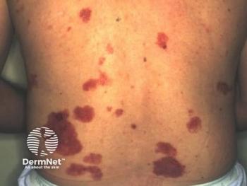
Palpable purpura
Regarding palpable purpura, approximately 10 percent of cases occurring in hospitalized patients are associated with drugs, Dr. Adams says.
“It’s probably a type 3 immune complex reaction. Antibiotics are at the top of the list of culprit drugs,” he says.
The generalized presentation of monomorphic symmetrical papules can signal systemic drug-associated acne, he says. It is almost always a reaction to steroids, he adds.
Somewhat similarly, he says, epidermal growth factor receptor (EGFR) inhibitors can cause a follicular eruption that shares features of acne and dermatitis.
“You can treat it like acne, but avoid irritants. Sometimes topical corticosteroids are needed for the dermatitis component,” he says.
Systemic medication reactions
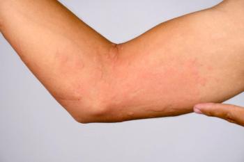
Additionally, Dr. Adams says patients commonly experience adverse reactions to various systemic medications (antihypertensives, hormones, anticholinergics and others) administered by an applied patch, which can cause erythematous patches within the applied contact surface or may extend beyond the dimensions of the original patch. If the reaction occurs beyond the patch border, some medication patch manufacturers recommend avoiding the systemic form of the drug to avoid a potential severe systemic reaction. All medication patches approved by the Food and Drug Administration can cause such a reaction, he states.
“It’s either allergic or irritant contact dermatitis. Rarely do these patients get seriously ill from it,” Dr. Adams says.
In one case that turned out to be allergic contact dermatitis, he says, a hospitalized patient had an epidermal catheter line taped from his back to his shoulder and the entire length of underlying skin “painted” with compound tincture of benzoin to improve tape adhesion. The patient later developed an intense pruritic reaction running the entire length of the catheter. The adhesive contained Tolu balsam, which Dr. Adams says is similar to and can cross-react with balsam of Peru (Myroxylon pereirae), an allergen often tested in patch test panels.
“Any time we use Tolu balsam to help tape or sterile strips adhere, there is potential for this kind of reaction,” he says.
Conversely, acral erythrodysesthesia is “probably related to a drug being excreted through the sweat glands and causing a local toxic reaction. These patients usually complain of a burning feeling of the hands and/or feet,” Dr. Adams says. After the medication is discontinued, “They usually heal satisfactorily. It is usually treated with local cold compresses.” Some reports have suggested topical or oral vitamin B6, he adds, “But I'm not sold on that.”
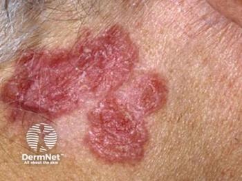
Highly pruritic flat-topped papules can represent lichen planus (LP)-like drug reaction, Dr. Adams says. In several cases, “Performing a biopsy helped us make the diagnosis. The key is, most often traditional LP affects the extremities, whereas drug-associated LP tends to impact the trunk.”
What makes this reaction difficult to diagnose is that it may not happen for months after the drug exposure, he adds.
“And when you stop the drug, it may take months to go away,” Dr. Adams says.
Disclosures: Dr. Adams reports no relevant financial interests.
Advertisement
Related Content
Advertisement


Latest CME


Advertisement
Advertisement
Trending on Dermatology Times
1
What Dermatologists Need to Know About the First New Sunscreen Ingredient in Decades
2
The Expanding Role of Immunotherapies for Non-Melanoma Skin Cancers
3
Once-Daily Zasocitinib Rivals Injectable Biologics for Skin Clearance, Phase 3 Data Show
4
Filling the HS Treatment Gap: Ruxolitinib Targets Early-Stage Disease
5