Compression sclerotherapy: Treatment of choice for leg veins
Dr. Goldman notes that postsclerosis compression is perhaps the most important advance in sclerotherapy for varicose veins since the introduction of synthetic sclerosing agents in the 1940s.
"Individually or in combination, varicose, reticular veins or telangiectatic veins appear in one-third of patients before age 25 and increase in incidence with age," says Mitchel P. Goldman, M.D., associate clinical professor of dermatology at the University of California, San Diego.
Definitions
To determine the best treatment option, he says, venous reflux needs to be considered first.
"Regardless of the underlying etiologic event leading to venous hypertension, the point of reflux - whether it is through either incompetent perforating veins, feeding reticular veins or an incompetent saphenofemoral junction (SFJ) - must be treated first," Dr. Goldman says, adding that several diagnostic tests can be performed to help define the extent of varicose vein disease and to plan treatment.
"The primary goal is to locate the point of high-pressure reflux, and Doppler ultrasound and duplex scanning are the major diagnostic techniques used," Dr. Goldman says. "The Doppler ultrasound is used to determine the flow of blood within the superficial veins. When placed over the clinically apparent varicose vein, flow can be heard when one compresses the vein distally. If blood is heard to flow distally, an incompetence of the venous valves is present."
Depending on the cause of the condition, surgery, sclerotherapy, laser or intense pulsed light or a combination of all techniques is necessary.
"Surgical ligation and stripping procedures are essentially procedures of the past," Dr. Goldman tells Dermatology Times. "For varicose veins more than 1 cm in diameter and for patients with an incompetent great saphenous vein, short saphenous vein and/or SFJ, I recommend endoluminal laser closure."
This technique is performed under local tumescent anesthesia, Dr. Goldman says, and the patient can walk out of the office immediately after the procedure.
"In our experience, the procedure causes no pain and patients can resume all normal activities within a day," he says. "I recommend the 1320 nm endoluminal laser, although other wavelengths and radiofrequency are also used."
Ambulatory phlebectomy
For veins 4 mm to 10 mm in diameter, Dr. Goldman recommends ambulatory phlebectomy.
"In this procedure, veins are removed through 2 mm to 3 mm incisions, and thus there is no chance of recurrence and there is a decreased risk of adverse sequelae," he says. "A beneficial effect of ambulatory phlebectomy is that the harvested veins can be transferred to areas that need filling in other procedures, such as the nasolabial grooves and lips, saving the patient who wants these procedures the cost of using temporary or artificial fillers."
Sclerotherapy
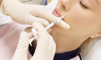
Dr. Goldman recommends sclerotherapy for veins less than 4 mm in diameter.
For veins 1 mm in diameter and/or for veins which are persistent after sclerotherapy and phlebectomy, laser or intense pulsed light treatment can also be used. He defines sclerotherapy as the introduction of a foreign substance into the lumen of a vessel, causing thrombosis and subsequent fibrosis.
Detergent sclerosants, such as sodium morrhuate, polidocanol and sodium tetradecyl sulfate produce endothelial damage through interference with the cell surface lipids. Hypertonic saline and hypertonic glucose solutions produce dehydration of endothelial cells through osmosis, resulting in endothelial destruction, while chemical irritants or caustic agents such as glycerin and polyiodinated iodine produce direct destruction of the endothelial cells.
Newsletter
Like what you’re reading? Subscribe to Dermatology Times for weekly updates on therapies, innovations, and real-world practice tips.


